 Movie
Movie Controller


+ Open data

- Basic information
| Entry | Database: PDB / ID: 1qsg | ||||||
|---|---|---|---|---|---|---|---|
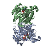
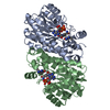
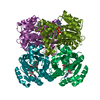
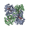
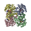
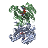
| Title | CRYSTAL STRUCTURE OF ENOYL REDUCTASE INHIBITION BY TRICLOSAN | ||||||
 Components Components | ENOYL-[ACYL-CARRIER-PROTEIN] REDUCTASE | ||||||
 Keywords Keywords | OXIDOREDUCTASE / ENOYL REDUCTASE | ||||||
| Function / homology |  Function and homology information Function and homology informationenoyl-[acyl-carrier-protein] reductase [NAD(P)H] activity / NADH binding / biotin biosynthetic process / enoyl-[acyl-carrier-protein] reductase (NADH) / fatty acid elongation / enoyl-[acyl-carrier-protein] reductase (NADH) activity / lipid biosynthetic process / catalytic complex / protein homotetramerization / response to antibiotic ...enoyl-[acyl-carrier-protein] reductase [NAD(P)H] activity / NADH binding / biotin biosynthetic process / enoyl-[acyl-carrier-protein] reductase (NADH) / fatty acid elongation / enoyl-[acyl-carrier-protein] reductase (NADH) activity / lipid biosynthetic process / catalytic complex / protein homotetramerization / response to antibiotic / protein-containing complex / membrane / identical protein binding / cytosol Similarity search - Function | ||||||
| Biological species |  | ||||||
| Method |  X-RAY DIFFRACTION / SYNCHROTRON / MOLECULAR REPLACEMENT / Resolution: 1.75 Å X-RAY DIFFRACTION / SYNCHROTRON / MOLECULAR REPLACEMENT / Resolution: 1.75 Å | ||||||
 Authors Authors | Stewart, M.J. / Parikh, S. / Xiao, G. / Tonge, P.J. / Kisker, C. | ||||||
 Citation Citation | Journal: J.Mol.Biol. / Year: 1999 Title: Structural basis and mechanism of enoyl reductase inhibition by triclosan. Authors: Stewart, M.J. / Parikh, S. / Xiao, G. / Tonge, P.J. / Kisker, C. | ||||||
| History |
|
- Structure visualization
| Structure viewer | Molecule: MolmilJmol/JSmol |
|---|
- Downloads & links
-Download
| PDBx/mmCIF format | 1qsg.cif.gz | 424.5 KB | Display | PDBx/mmCIF format |
|---|---|---|---|---|
| PDB format | pdb1qsg.ent.gz | 345.7 KB | Display | PDB format |
| PDBx/mmJSON format | 1qsg.json.gz | Tree view | PDBx/mmJSON format | |
| Others |  Other downloads Other downloads |
-Validation report
| Arichive directory | https://data.pdbj.org/pub/pdb/validation_reports/qs/1qsgftp://data.pdbj.org/pub/pdb/validation_reports/qs/1qsg | HTTPS FTP |
|---|
-Related structure data
| Related structure data |  1dfiS S: Starting model for refinement |
|---|---|
| Similar structure data |


-Links
 PDBj
PDBj


- Assembly
| Deposited unit | 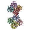
| ||||||||
|---|---|---|---|---|---|---|---|---|---|
| 1 | 
| ||||||||
| 2 | 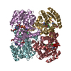
| ||||||||
| Unit cell |
|
-Components
| #1: Protein | Mass: 28175.201 Da / Num. of mol.: 8 Source method: isolated from a genetically manipulated source Source: (gene. exp.) References: UniProt: P29132, UniProt: P0AEK4*PLUS, enoyl-[acyl-carrier-protein] reductase (NADH) #2: Sugar | ChemComp-GLC /   Type: D-saccharide, alpha linking / Mass: 180.156 Da / Num. of mol.: 8 Type: D-saccharide, alpha linking / Mass: 180.156 Da / Num. of mol.: 8Source method: isolated from a genetically manipulated source Formula: C6H12O6 #3: Chemical | ChemComp-NAD /   Mass: 663.425 Da / Num. of mol.: 8 / Source method: obtained synthetically / Formula: C21H27N7O14P2 / Comment: NAD*YM Mass: 663.425 Da / Num. of mol.: 8 / Source method: obtained synthetically / Formula: C21H27N7O14P2 / Comment: NAD*YM#4: Chemical | ChemComp-TCL /   Mass: 289.542 Da / Num. of mol.: 8 / Source method: obtained synthetically / Formula: C12H7Cl3O2 / Comment: antifungal, antibiotic, detergent*YM Mass: 289.542 Da / Num. of mol.: 8 / Source method: obtained synthetically / Formula: C12H7Cl3O2 / Comment: antifungal, antibiotic, detergent*YM#5: Water | ChemComp-HOH / |  Mass: 18.015 Da / Num. of mol.: 1350 / Source method: isolated from a natural source / Formula: H2O Mass: 18.015 Da / Num. of mol.: 1350 / Source method: isolated from a natural source / Formula: H2O |
|---|
-Experimental details
-Experiment
| Experiment | Method: X-RAY DIFFRACTION / Number of used crystals: 1 |
|---|
- Sample preparation
| Crystal | Density Matthews: 2.2 Å3/Da / Density % sol: 43 % |
|---|---|
| Crystal grow | Temperature: 295.15 K / Method: vapor diffusion, hanging drop / pH: 4.6 Details: PEG 4000, AMMONIUM ACETATE, SODIUM ACETATE, pH 4.6, VAPOR DIFFUSION, HANGING DROP, temperature 295.15K |
-Data collection
| Diffraction | Mean temperature: 95 K |
|---|---|
| Diffraction source | Source: SYNCHROTRON / Site: NSLS  / Beamline: X26C / Wavelength: 1.1 / Beamline: X26C / Wavelength: 1.1 |
| Detector | Type: ADSC QUANTUM 4r / Detector: CCD / Date: Feb 7, 1999 |
| Radiation | Protocol: SINGLE WAVELENGTH / Monochromatic (M) / Laue (L): M / Scattering type: x-ray |
| Radiation wavelength | Wavelength: 1.1 Å / Relative weight: 1 |
| Reflection | Resolution: 1.75→20 Å / Num. obs: 183224 / % possible obs: 94.2 % / Observed criterion σ(I): 0 / Redundancy: 1.85 % / Biso Wilson estimate: 24.7 Å2 / Rmerge(I) obs: 0.044 / Net I/σ(I): 13.2 |
| Reflection shell | Resolution: 1.75→1.81 Å / Redundancy: 1.64 % / Rmerge(I) obs: 0.357 / Mean I/σ(I) obs: 1.6 / % possible all: 90.5 |
| Reflection shell | *PLUS % possible obs: 90.5 % |
- Processing
| Software |
| |||||||||||||||||||||||||||||||||||||||||||||||||||||||||||||||
|---|---|---|---|---|---|---|---|---|---|---|---|---|---|---|---|---|---|---|---|---|---|---|---|---|---|---|---|---|---|---|---|---|---|---|---|---|---|---|---|---|---|---|---|---|---|---|---|---|---|---|---|---|---|---|---|---|---|---|---|---|---|---|---|---|
| Refinement | Method to determine structure: MOLECULAR REPLACEMENT Starting model: PDB ENTRY 1DFI Resolution: 1.75→20 Å / SU B: 3.26 / SU ML: 0.1 / Cross valid method: THROUGHOUT / σ(F): 0 / ESU R: 0.12 / ESU R Free: 0.12
| |||||||||||||||||||||||||||||||||||||||||||||||||||||||||||||||
| Displacement parameters | Biso mean: 27.18 Å2 | |||||||||||||||||||||||||||||||||||||||||||||||||||||||||||||||
| Refinement step | Cycle: LAST / Resolution: 1.75→20 Å
| |||||||||||||||||||||||||||||||||||||||||||||||||||||||||||||||
| Refine LS restraints |
|
