 Movie
Movie Controller

[English] 日本語
 Yorodumi
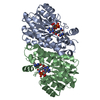
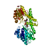
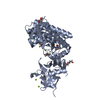
- PDB-4jx8: Crystal Structure of E.coli Enoyl Reductase in Complex with NAD a... -
+ Open data

- Basic information
| Entry | Database: PDB / ID: 4jx8 | ||||||
|---|---|---|---|---|---|---|---|
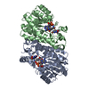
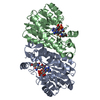
| Title | Crystal Structure of E.coli Enoyl Reductase in Complex with NAD and AEA16 | ||||||
 Components Components | Enoyl-[acyl-carrier-protein] reductase [NADH] FabI | ||||||
 Keywords Keywords | OXIDOREDUCTASE/OXIDOREDUCTASE INHIBITOR / Fabi / Ligand AEA16 / Enoyl Reductase / OXIDOREDUCTASE-OXIDOREDUCTASE INHIBITOR complex | ||||||
| Function / homology |  Function and homology information Function and homology informationNADH binding / biotin biosynthetic process / enoyl-[acyl-carrier-protein] reductase (NADH) / fatty acid elongation / enoyl-[acyl-carrier-protein] reductase (NADH) activity / lipid biosynthetic process / catalytic complex / protein homotetramerization / response to antibiotic / protein-containing complex ...NADH binding / biotin biosynthetic process / enoyl-[acyl-carrier-protein] reductase (NADH) / fatty acid elongation / enoyl-[acyl-carrier-protein] reductase (NADH) activity / lipid biosynthetic process / catalytic complex / protein homotetramerization / response to antibiotic / protein-containing complex / membrane / identical protein binding / cytosol Similarity search - Function | ||||||
| Biological species |  | ||||||
| Method |  X-RAY DIFFRACTION / MOLECULAR REPLACEMENT / Resolution: 3.2 Å X-RAY DIFFRACTION / MOLECULAR REPLACEMENT / Resolution: 3.2 Å | ||||||
 Authors Authors | Subramanya, H. / Rao, K.N. / Anirudha, L. | ||||||
 Citation Citation | Journal: To be Published Title: Crystal Structure of E.coli Enoyl Reductase in Complex with NAD and AEA16 Authors: Subramanya, H. / Rao, K.N. / Anirudha, L. | ||||||
| History |
|
- Structure visualization
| Structure viewer | Molecule: MolmilJmol/JSmol |
|---|
- Downloads & links
-Download
| PDBx/mmCIF format | 4jx8.cif.gz | 111.1 KB | Display | PDBx/mmCIF format |
|---|---|---|---|---|
| PDB format | pdb4jx8.ent.gz | 85.2 KB | Display | PDB format |
| PDBx/mmJSON format | 4jx8.json.gz | Tree view | PDBx/mmJSON format | |
| Others |  Other downloads Other downloads |
-Validation report
| Arichive directory | https://data.pdbj.org/pub/pdb/validation_reports/jx/4jx8ftp://data.pdbj.org/pub/pdb/validation_reports/jx/4jx8 | HTTPS FTP |
|---|
-Related structure data
| Related structure data | 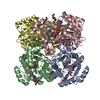 1qsgS S: Starting model for refinement |
|---|---|
| Similar structure data |


-Links
 PDBj
PDBj

- Assembly
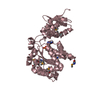
| Deposited unit | 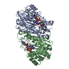
| ||||||||
|---|---|---|---|---|---|---|---|---|---|
| 1 |
| ||||||||
| Unit cell |
|
-Components
| #1: Protein | Mass: 30064.279 Da / Num. of mol.: 2 Source method: isolated from a genetically manipulated source Source: (gene. exp.) References: UniProt: P0AEK4, enoyl-[acyl-carrier-protein] reductase (NADH) #2: Chemical |   Mass: 385.415 Da / Num. of mol.: 2 / Source method: obtained synthetically / Formula: C23H19N3O3 Mass: 385.415 Da / Num. of mol.: 2 / Source method: obtained synthetically / Formula: C23H19N3O3#3: Chemical |   Mass: 663.425 Da / Num. of mol.: 2 / Source method: obtained synthetically / Formula: C21H27N7O14P2 / Comment: NAD*YM Mass: 663.425 Da / Num. of mol.: 2 / Source method: obtained synthetically / Formula: C21H27N7O14P2 / Comment: NAD*YM#4: Water | ChemComp-HOH / |  Mass: 18.015 Da / Num. of mol.: 54 / Source method: isolated from a natural source / Formula: H2O Mass: 18.015 Da / Num. of mol.: 54 / Source method: isolated from a natural source / Formula: H2O |
|---|
-Experimental details
-Experiment
| Experiment | Method: X-RAY DIFFRACTION / Number of used crystals: 1 |
|---|
- Sample preparation
| Crystal | Density Matthews: 2.46 Å3/Da / Density % sol: 50.02 % |
|---|---|
| Crystal grow | Temperature: 277 K / Method: vapor diffusion, hanging drop / pH: 4.6 Details: 0.1M Sodium acetate pH 4.6, 10%-15% (w/v) PEG 4000, 200mM Ammonium acetate, VAPOR DIFFUSION, HANGING DROP, temperature 277K |
-Data collection
| Diffraction | Mean temperature: 100 K |
|---|---|
| Diffraction source | Source: ROTATING ANODE / Type: RIGAKU RU300 / Wavelength: 1.5418 Å |
| Detector | Type: RIGAKU RAXIS IV++ / Detector: IMAGE PLATE / Details: Mirrors |
| Radiation | Monochromator: Ni fliter / Protocol: SINGLE WAVELENGTH / Monochromatic (M) / Laue (L): M / Scattering type: x-ray |
| Radiation wavelength | Wavelength: 1.5418 Å / Relative weight: 1 |
| Reflection | Resolution: 3.2→30 Å / Num. obs: 8204 / % possible obs: 81.3 % / Redundancy: 6.6 % / Rmerge(I) obs: 0.092 / Net I/σ(I): 37.5 |
| Reflection shell | Resolution: 3.2→3.31 Å / Redundancy: 5.8 % / Rmerge(I) obs: 0.137 / Mean I/σ(I) obs: 4.5 / Num. unique all: 811 / % possible all: 84.6 |
- Processing
| Software |
| |||||||||||||||||||||||||||||||||||||||||||||
|---|---|---|---|---|---|---|---|---|---|---|---|---|---|---|---|---|---|---|---|---|---|---|---|---|---|---|---|---|---|---|---|---|---|---|---|---|---|---|---|---|---|---|---|---|---|---|
| Refinement | Method to determine structure: MOLECULAR REPLACEMENT Starting model: 1QSG Resolution: 3.2→29.24 Å / Cor.coef. Fo:Fc: 0.879 / Cor.coef. Fo:Fc free: 0.702 / SU B: 28.243 / SU ML: 0.477 / Cross valid method: THROUGHOUT / ESU R Free: 0.819 / Stereochemistry target values: MAXIMUM LIKELIHOOD / Details: HYDROGENS HAVE BEEN USED IF PRESENT IN THE INPUT
| |||||||||||||||||||||||||||||||||||||||||||||
| Solvent computation | Ion probe radii: 0.8 Å / Shrinkage radii: 0.8 Å / VDW probe radii: 1.2 Å / Solvent model: MASK | |||||||||||||||||||||||||||||||||||||||||||||
| Displacement parameters | Biso mean: 24.295 Å2
| |||||||||||||||||||||||||||||||||||||||||||||
| Refinement step | Cycle: LAST / Resolution: 3.2→29.24 Å
| |||||||||||||||||||||||||||||||||||||||||||||
| Refine LS restraints |
| |||||||||||||||||||||||||||||||||||||||||||||
| LS refinement shell | Resolution: 3.2→3.282 Å / Total num. of bins used: 20
|
