 Movie
Movie Controller

[English] 日本語
 Yorodumi
- PDB-4aem: Structural and biochemical characterization of a novel Carbohydra... -
+ Open data

- Basic information
| Entry | Database: PDB / ID: 4aem | ||||||
|---|---|---|---|---|---|---|---|
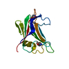
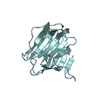
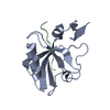
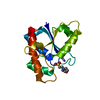
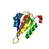
| Title | Structural and biochemical characterization of a novel Carbohydrate Binding Module of endoglucanase Cel5A from Eubacterium cellulosolvens | ||||||
 Components Components | ENDOGLUCANASE CEL5A | ||||||
 Keywords Keywords | HYDROLASE / FAMILY 5 GLYCOSIDE HYDROLASE / CELLULOSOME | ||||||
| Function / homology |  Function and homology information Function and homology informationbeta-glucosidase activity / cellulose catabolic process / cell surface / extracellular region Similarity search - Function | ||||||
| Biological species |  EUBACTERIUM CELLULOSOLVENS (bacteria) EUBACTERIUM CELLULOSOLVENS (bacteria) | ||||||
| Method |  X-RAY DIFFRACTION / SYNCHROTRON / MOLECULAR REPLACEMENT / Resolution: 2.1 Å X-RAY DIFFRACTION / SYNCHROTRON / MOLECULAR REPLACEMENT / Resolution: 2.1 Å | ||||||
 Authors Authors | Luis, A.S. / Venditto, I. / Prates, J.A.M. / Ferreira, L.M.A. / Gilbert, H.J. / Fontes, C.M.G.A. / Najmudin, S. | ||||||
 Citation Citation | Journal: J.Biol.Chem. / Year: 2013 Title: Understanding How Non-Catalytic Carbohydrate Binding Modules Can Display Specificity for Xyloglucan Authors: Luis, A.S. / Venditto, I. / Prates, J.A.M. / Ferreira, L.M.A. / Temple, M.J. / Rogowski, A. / Basle, A. / Xue, J. / Knox, J.P. / Najmudin, S. / Fontes, C.M.G.A. / Gilbert, H.J. #1: Journal: Acta Crystallogr.,Sect.F / Year: 2011 Title: Overproduction, Purification, Crystallization and Preliminary X-Ray Characterization of a Novel Carbohydrate-Binding Module of Endoglucanase Cel5A from Eubacterium Cellulosolvens. Authors: Luis, A.S. / Alves, V.D. / Romao, M.J. / Prates, J.A.M. / Fontes, C.M.G.A. / Najmudin, S. | ||||||
| History |
|
- Structure visualization
| Structure viewer | Molecule: MolmilJmol/JSmol |
|---|
- Downloads & links
-Download
| PDBx/mmCIF format | 4aem.cif.gz | 66.3 KB | Display | PDBx/mmCIF format |
|---|---|---|---|---|
| PDB format | pdb4aem.ent.gz | 49.7 KB | Display | PDB format |
| PDBx/mmJSON format | 4aem.json.gz | Tree view | PDBx/mmJSON format | |
| Others |  Other downloads Other downloads |
-Validation report
| Arichive directory | https://data.pdbj.org/pub/pdb/validation_reports/ae/4aemftp://data.pdbj.org/pub/pdb/validation_reports/ae/4aem | HTTPS FTP |
|---|
-Related structure data
| Related structure data |  2ypjC  4aekSC  4afdC  4afmC 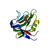 4ba6C S: Starting model for refinement C: citing same article ( |
|---|---|
| Similar structure data |


-Links
 PDBj
PDBj- Assembly
| Deposited unit | 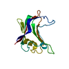
| ||||||||
|---|---|---|---|---|---|---|---|---|---|
| 1 |
| ||||||||
| Unit cell |
|
-Components
| #1: Protein | Mass: 14799.314 Da / Num. of mol.: 1 / Fragment: CARBOHYDRATE BINDING MODULE, RESIDUES 37-170 Source method: isolated from a genetically manipulated source Source: (gene. exp.) EUBACTERIUM CELLULOSOLVENS (bacteria) / Production host: |
|---|---|
| #2: Water | ChemComp-HOH /  Mass: 18.015 Da / Num. of mol.: 92 / Source method: isolated from a natural source / Formula: H2O Mass: 18.015 Da / Num. of mol.: 92 / Source method: isolated from a natural source / Formula: H2O |
-Experimental details
-Experiment
| Experiment | Method: X-RAY DIFFRACTION / Number of used crystals: 1 |
|---|
- Sample preparation
| Crystal | Density Matthews: 2.32 Å3/Da / Density % sol: 47 % / Description: NONE |
|---|---|
| Crystal grow | Temperature: 292 K / pH: 4.76 Details: 100 MG/ML PROTEIN WAS USED AT 292 K WITH 0.2 M AMMONIUM SULFATE, 0.1 M SODIUM ACETATE TRIHYDRATE PH 4.6, 30% W/V PEG 2K MME. 30% GLYCEROL WAS USED AS CRYOPROTECTANT |
-Data collection
| Diffraction | Mean temperature: 100 K |
|---|---|
| Diffraction source | Source: SYNCHROTRON / Site: ESRF  / Beamline: ID14-4 / Wavelength: 0.9793 / Beamline: ID14-4 / Wavelength: 0.9793 |
| Detector | Type: ADSC QUANTUM 315r / Detector: CCD / Date: Jun 24, 2011 |
| Radiation | Protocol: SINGLE WAVELENGTH / Monochromatic (M) / Laue (L): M / Scattering type: x-ray |
| Radiation wavelength | Wavelength: 0.9793 Å / Relative weight: 1 |
| Reflection | Resolution: 2.1→68.69 Å / Num. obs: 8165 / % possible obs: 83.6 % / Redundancy: 11.2 % / Rmerge(I) obs: 0.11 / Net I/σ(I): 15.3 |
| Reflection shell | Resolution: 2.1→2.21 Å / Redundancy: 6.8 % / Rmerge(I) obs: 0.8 / Mean I/σ(I) obs: 2.4 / % possible all: 53.4 |
- Processing
| Software |
| ||||||||||||||||||||||||||||||||||||||||||||||||||||||||||||||||||||||||||||||||||||||||||||||||||||||||||||||||||||||||||||||||||||||||||||||||||||||||||||||||||||||||||||||||||||||
|---|---|---|---|---|---|---|---|---|---|---|---|---|---|---|---|---|---|---|---|---|---|---|---|---|---|---|---|---|---|---|---|---|---|---|---|---|---|---|---|---|---|---|---|---|---|---|---|---|---|---|---|---|---|---|---|---|---|---|---|---|---|---|---|---|---|---|---|---|---|---|---|---|---|---|---|---|---|---|---|---|---|---|---|---|---|---|---|---|---|---|---|---|---|---|---|---|---|---|---|---|---|---|---|---|---|---|---|---|---|---|---|---|---|---|---|---|---|---|---|---|---|---|---|---|---|---|---|---|---|---|---|---|---|---|---|---|---|---|---|---|---|---|---|---|---|---|---|---|---|---|---|---|---|---|---|---|---|---|---|---|---|---|---|---|---|---|---|---|---|---|---|---|---|---|---|---|---|---|---|---|---|---|---|
| Refinement | Method to determine structure: MOLECULAR REPLACEMENT Starting model: PDB ENTRY 4AEK Resolution: 2.1→68.69 Å / Cor.coef. Fo:Fc: 0.962 / Cor.coef. Fo:Fc free: 0.931 / SU B: 10.403 / SU ML: 0.132 / Cross valid method: THROUGHOUT / ESU R: 0.223 / ESU R Free: 0.194 / Stereochemistry target values: MAXIMUM LIKELIHOOD Details: HYDROGENS HAVE BEEN ADDED IN THE RIDING POSITIONS. HYDROGENS HAVE BEEN USED IF PRESENT IN THE INPUT. PHENIX-1.7.2-869 WAS ALSO USED FOR REFINEMENT, TO GENERATE TLS GROUPS AND UPDATE WATERS. ...Details: HYDROGENS HAVE BEEN ADDED IN THE RIDING POSITIONS. HYDROGENS HAVE BEEN USED IF PRESENT IN THE INPUT. PHENIX-1.7.2-869 WAS ALSO USED FOR REFINEMENT, TO GENERATE TLS GROUPS AND UPDATE WATERS. RESIDUES A37-38 AND A165-170 ARE DISORDERERED. GLU 65, ILE 111, AND GLU 126 WERE GIVEN DUAL CONFORMATIONS. DISORDERED SIDE CHAINS WERE MODELED STEREOCHEMICALLY.
| ||||||||||||||||||||||||||||||||||||||||||||||||||||||||||||||||||||||||||||||||||||||||||||||||||||||||||||||||||||||||||||||||||||||||||||||||||||||||||||||||||||||||||||||||||||||
| Solvent computation | Ion probe radii: 0.8 Å / Shrinkage radii: 0.8 Å / VDW probe radii: 1.2 Å / Solvent model: MASK | ||||||||||||||||||||||||||||||||||||||||||||||||||||||||||||||||||||||||||||||||||||||||||||||||||||||||||||||||||||||||||||||||||||||||||||||||||||||||||||||||||||||||||||||||||||||
| Displacement parameters | Biso mean: 31.376 Å2
| ||||||||||||||||||||||||||||||||||||||||||||||||||||||||||||||||||||||||||||||||||||||||||||||||||||||||||||||||||||||||||||||||||||||||||||||||||||||||||||||||||||||||||||||||||||||
| Refinement step | Cycle: LAST / Resolution: 2.1→68.69 Å
| ||||||||||||||||||||||||||||||||||||||||||||||||||||||||||||||||||||||||||||||||||||||||||||||||||||||||||||||||||||||||||||||||||||||||||||||||||||||||||||||||||||||||||||||||||||||
| Refine LS restraints |
|