 Movie
Movie Controller

[English] 日本語
 Yorodumi
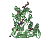
- PDB-4afm: Structural and biochemical characterization of a novel Carbohydra... -
+ Open data

- Basic information
| Entry | Database: PDB / ID: 4afm | ||||||
|---|---|---|---|---|---|---|---|
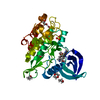
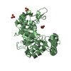
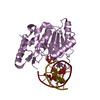
| Title | Structural and biochemical characterization of a novel Carbohydrate Binding Module of endoglucanase Cel5A from Eubacterium cellulosolvens. | ||||||
 Components Components | ENDOGLUCANASE CEL5A | ||||||
 Keywords Keywords | HYDROLASE / CARBOHYDRATE BINDING MODULE / FAMILY 5 GLYCOSIDE HYDROLASE | ||||||
| Function / homology |  Function and homology information Function and homology informationbeta-glucosidase activity / cellulose catabolic process / cell surface / extracellular region Similarity search - Function | ||||||
| Biological species |  EUBACTERIUM CELLULOSOLVENS (bacteria) EUBACTERIUM CELLULOSOLVENS (bacteria) | ||||||
| Method |  X-RAY DIFFRACTION / SYNCHROTRON / MOLECULAR REPLACEMENT / Resolution: 1.25 Å X-RAY DIFFRACTION / SYNCHROTRON / MOLECULAR REPLACEMENT / Resolution: 1.25 Å | ||||||
 Authors Authors | Luis, A.S. / Venditto, I. / Prates, J.A.M. / Ferreira, L.M.A. / Gilbert, H.J. / Fontes, C.M.G.A. / Najmudin, S. | ||||||
 Citation Citation | Journal: J.Biol.Chem. / Year: 2013 Title: Understanding How Non-Catalytic Carbohydrate Binding Modules Can Display Specificity for Xyloglucan. Authors: Luis, A.S. / Venditto, I. / Prates, J.A.M. / Ferrieira, L.M.A. / Temple, M.J. / Rogowski, A. / Basle, A. / Xue, J. / Knox, J.P. / Najmudin, S. / Fontes, C.M.G.A. / Gilbert, H.J. #1: Journal: Acta Crystallogr.,Sect.F / Year: 2011 Title: Overproduction, Purification, Crystallization and Preliminary X-Ray Characterization of a Novel Carbohydrate-Binding Module of Endoglucanase Cel5A from Eubacterium Cellulosolvens. Authors: Luis, A.S. / Alves, V.D. / Romao, M.J. / Prates, J.A.M. / Fontes, C.M.G.A. / Najmudin, S. | ||||||
| History |
|
- Structure visualization
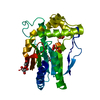
| Structure viewer | Molecule: MolmilJmol/JSmol |
|---|
- Downloads & links
-Download
| PDBx/mmCIF format | 4afm.cif.gz | 72 KB | Display | PDBx/mmCIF format |
|---|---|---|---|---|
| PDB format | pdb4afm.ent.gz | 54.3 KB | Display | PDB format |
| PDBx/mmJSON format | 4afm.json.gz | Tree view | PDBx/mmJSON format | |
| Others |  Other downloads Other downloads |
-Validation report
| Arichive directory | https://data.pdbj.org/pub/pdb/validation_reports/af/4afmftp://data.pdbj.org/pub/pdb/validation_reports/af/4afm | HTTPS FTP |
|---|
-Related structure data
| Related structure data |  2ypjC  4aekSC 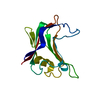 4aemC  4afdC 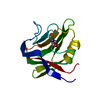 4ba6C C: citing same article ( S: Starting model for refinement |
|---|---|
| Similar structure data |


-Links
 PDBj
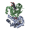
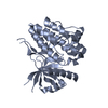
PDBj- Assembly
| Deposited unit | 
| |||||||||
|---|---|---|---|---|---|---|---|---|---|---|
| 1 | 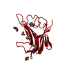
| |||||||||
| Unit cell |
| |||||||||
| Components on special symmetry positions |
|
-Components
| #1: Protein | Mass: 14879.080 Da / Num. of mol.: 1 / Fragment: CARBOHYDRATE BINDING MODULE, RESIDUES 37-170 Source method: isolated from a genetically manipulated source Source: (gene. exp.) EUBACTERIUM CELLULOSOLVENS (bacteria) / Production host: | ||||||||
|---|---|---|---|---|---|---|---|---|---|
| #2: Chemical |   Mass: 92.094 Da / Num. of mol.: 3 / Source method: obtained synthetically / Formula: C3H8O3 Mass: 92.094 Da / Num. of mol.: 3 / Source method: obtained synthetically / Formula: C3H8O3#3: Chemical | ChemComp-ACT /   Mass: 59.044 Da / Num. of mol.: 4 / Source method: obtained synthetically / Formula: C2H3O2 Mass: 59.044 Da / Num. of mol.: 4 / Source method: obtained synthetically / Formula: C2H3O2#4: Water | ChemComp-HOH / |  Mass: 18.015 Da / Num. of mol.: 130 / Source method: isolated from a natural source / Formula: H2O Mass: 18.015 Da / Num. of mol.: 130 / Source method: isolated from a natural source / Formula: H2OHas protein modification | Y | Nonpolymer details | GLYCEROL (GOL): FROM THE CRYOPROTEC | |
-Experimental details
-Experiment
| Experiment | Method: X-RAY DIFFRACTION / Number of used crystals: 1 |
|---|
- Sample preparation
| Crystal | Density Matthews: 3.65 Å3/Da / Density % sol: 66 % / Description: NONE |
|---|---|
| Crystal grow | Temperature: 292 K / pH: 4.6 Details: 80 MG/ML OF PROTEIN AT 292 K WERE GROWN IN 0.2 M AMMONIUM SULFATE, 0.1 M SODIUM ACETATE TRIHYDRATE PH 4.6, 26% W/V PEG 2K MME. CRYSTALS WERE SOAKED WITH 10 MM CELLOHEXAOSE FOR A FEW HOURS. ...Details: 80 MG/ML OF PROTEIN AT 292 K WERE GROWN IN 0.2 M AMMONIUM SULFATE, 0.1 M SODIUM ACETATE TRIHYDRATE PH 4.6, 26% W/V PEG 2K MME. CRYSTALS WERE SOAKED WITH 10 MM CELLOHEXAOSE FOR A FEW HOURS. 30% GLYCEROL WAS USED AS A CRYOPROTECTANT. |
-Data collection
| Diffraction | Mean temperature: 100 K |
|---|---|
| Diffraction source | Source: SYNCHROTRON / Site: SOLEIL  / Beamline: PROXIMA 1 / Wavelength: 0.97934 / Beamline: PROXIMA 1 / Wavelength: 0.97934 |
| Detector | Type: ADSC QUANTUM 315r / Detector: CCD / Date: Apr 4, 2011 |
| Radiation | Protocol: SINGLE WAVELENGTH / Monochromatic (M) / Laue (L): M / Scattering type: x-ray |
| Radiation wavelength | Wavelength: 0.97934 Å / Relative weight: 1 |
| Reflection | Resolution: 1.25→38.74 Å / Num. obs: 35557 / % possible obs: 91.2 % / Redundancy: 21.6 % / Rmerge(I) obs: 0.07 / Net I/σ(I): 28.2 |
| Reflection shell | Resolution: 1.25→1.32 Å / Redundancy: 6.2 % / Rmerge(I) obs: 0.4 / Mean I/σ(I) obs: 4 / % possible all: 62.4 |
- Processing
| Software |
| ||||||||||||||||||||||||||||||||||||||||||||||||||||||||||||||||||||||||||||||||||||||||||||||||||||||||||||||||||||||||||||||||||||||||||||||||||||||||||||||||||||||||||||||||||||||
|---|---|---|---|---|---|---|---|---|---|---|---|---|---|---|---|---|---|---|---|---|---|---|---|---|---|---|---|---|---|---|---|---|---|---|---|---|---|---|---|---|---|---|---|---|---|---|---|---|---|---|---|---|---|---|---|---|---|---|---|---|---|---|---|---|---|---|---|---|---|---|---|---|---|---|---|---|---|---|---|---|---|---|---|---|---|---|---|---|---|---|---|---|---|---|---|---|---|---|---|---|---|---|---|---|---|---|---|---|---|---|---|---|---|---|---|---|---|---|---|---|---|---|---|---|---|---|---|---|---|---|---|---|---|---|---|---|---|---|---|---|---|---|---|---|---|---|---|---|---|---|---|---|---|---|---|---|---|---|---|---|---|---|---|---|---|---|---|---|---|---|---|---|---|---|---|---|---|---|---|---|---|---|---|
| Refinement | Method to determine structure: MOLECULAR REPLACEMENT Starting model: PDB ENTRY 4AEK Resolution: 1.25→38.7 Å / Cor.coef. Fo:Fc: 0.967 / Cor.coef. Fo:Fc free: 0.963 / SU B: 1.074 / SU ML: 0.023 / Cross valid method: THROUGHOUT / ESU R: 0.043 / ESU R Free: 0.044 / Stereochemistry target values: MAXIMUM LIKELIHOOD Details: HYDROGENS HAVE BEEN ADDED IN THE RIDING POSITIONS. HYDROGENS HAVE BEEN USED IF PRESENT IN THE INPUT U VALUES.PHENIX-1.7.2-869 WAS USED IN THE PENULTIMATE STEP FOR OCCUPANCY REFINEMENT AND ...Details: HYDROGENS HAVE BEEN ADDED IN THE RIDING POSITIONS. HYDROGENS HAVE BEEN USED IF PRESENT IN THE INPUT U VALUES.PHENIX-1.7.2-869 WAS USED IN THE PENULTIMATE STEP FOR OCCUPANCY REFINEMENT AND TLS GROUP DETERMINATION. RESIDUES 37-38 AND 166-170 ARE DISORDERED. ASP A118 WAS MODELLED WITH ALTERNATE CONFORMATION. IT MAY ALSO HAVE RADIATION DAMAGE AS IN PDB 4AFD. DISORDERED SIDE CHAINS WERE MODELED STEREOCHEMICALLY. FOLLOWING WERE GIVEN ALTERNATE CONFORMATIONS, SER A45, GLU A51, GLN A67, GLN A71, MSE A73, GLU A82, ILE A83, IL3 A95 GLU A100, SER A103, SER A112, VAL A116, GLN A126, LYS A130, MSE A154, VAL A160 AND SER A162.
| ||||||||||||||||||||||||||||||||||||||||||||||||||||||||||||||||||||||||||||||||||||||||||||||||||||||||||||||||||||||||||||||||||||||||||||||||||||||||||||||||||||||||||||||||||||||
| Solvent computation | Ion probe radii: 0.8 Å / Shrinkage radii: 0.8 Å / VDW probe radii: 1.2 Å / Solvent model: MASK | ||||||||||||||||||||||||||||||||||||||||||||||||||||||||||||||||||||||||||||||||||||||||||||||||||||||||||||||||||||||||||||||||||||||||||||||||||||||||||||||||||||||||||||||||||||||
| Displacement parameters | Biso mean: 14.031 Å2
| ||||||||||||||||||||||||||||||||||||||||||||||||||||||||||||||||||||||||||||||||||||||||||||||||||||||||||||||||||||||||||||||||||||||||||||||||||||||||||||||||||||||||||||||||||||||
| Refinement step | Cycle: LAST / Resolution: 1.25→38.7 Å
| ||||||||||||||||||||||||||||||||||||||||||||||||||||||||||||||||||||||||||||||||||||||||||||||||||||||||||||||||||||||||||||||||||||||||||||||||||||||||||||||||||||||||||||||||||||||
| Refine LS restraints |
|