 Movie
Movie Controller


+ Open data

- Basic information
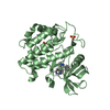
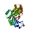
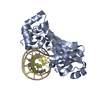
| Entry | Database: PDB / ID: 1hl6 | ||||||
|---|---|---|---|---|---|---|---|
| Title | A novel mode of RBD-protein recognition in the Y14-mago complex | ||||||
 Components Components |
| ||||||
 Keywords Keywords | SIGNAL PROTEIN / RDB / EXON-EXON JUNCTION / OSKAR / RNP / NMD | ||||||
| Function / homology |  Function and homology information Function and homology informationestablishment of pole plasm mRNA localization / oocyte nucleus localization involved in oocyte dorsal/ventral axis specification / maintenance of pole plasm mRNA location / Transport of Mature mRNA derived from an Intron-Containing Transcript / mRNA 3'-end processing / RNA Polymerase II Transcription Termination / Nonsense Mediated Decay (NMD) enhanced by the Exon Junction Complex (EJC) / oocyte anterior/posterior axis specification / mRNA Splicing - Major Pathway / pole plasm ...establishment of pole plasm mRNA localization / oocyte nucleus localization involved in oocyte dorsal/ventral axis specification / maintenance of pole plasm mRNA location / Transport of Mature mRNA derived from an Intron-Containing Transcript / mRNA 3'-end processing / RNA Polymerase II Transcription Termination / Nonsense Mediated Decay (NMD) enhanced by the Exon Junction Complex (EJC) / oocyte anterior/posterior axis specification / mRNA Splicing - Major Pathway / pole plasm / regulation of pole plasm oskar mRNA localization / oocyte dorsal/ventral axis specification / pole plasm assembly / oocyte microtubule cytoskeleton polarization / oocyte microtubule cytoskeleton organization / germarium-derived oocyte fate determination / pole plasm oskar mRNA localization / exon-exon junction complex / import into nucleus / NLS-dependent protein nuclear import complex / precatalytic spliceosome / oogenesis / mRNA transport / catalytic step 2 spliceosome / RNA splicing / mRNA splicing, via spliceosome / microtubule cytoskeleton organization / intracellular protein localization / mRNA binding / positive regulation of gene expression / RNA binding / nucleus / cytoplasm Similarity search - Function | ||||||
| Biological species |  | ||||||
| Method |  X-RAY DIFFRACTION / SYNCHROTRON / MIR / Resolution: 2.5 Å X-RAY DIFFRACTION / SYNCHROTRON / MIR / Resolution: 2.5 Å | ||||||
 Authors Authors | Fribourg, S. / Gatfield, D. / Yao, W. / Izaurralde, E. / Conti, E. | ||||||
 Citation Citation | Journal: Nat.Struct.Biol. / Year: 2003 Title: A Novel Mode of Rbd-Protein Recognition in the Y14-Mago Complex Authors: Fribourg, S. / Gatfield, D. / Izaurralde, E. / Conti, E. | ||||||
| History |
|
- Structure visualization
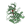
| Structure viewer | Molecule: MolmilJmol/JSmol |
|---|
- Downloads & links
-Download
| PDBx/mmCIF format | 1hl6.cif.gz | 113.6 KB | Display | PDBx/mmCIF format |
|---|---|---|---|---|
| PDB format | pdb1hl6.ent.gz | 90.2 KB | Display | PDB format |
| PDBx/mmJSON format | 1hl6.json.gz | Tree view | PDBx/mmJSON format | |
| Others |  Other downloads Other downloads |
-Validation report
| Arichive directory | https://data.pdbj.org/pub/pdb/validation_reports/hl/1hl6ftp://data.pdbj.org/pub/pdb/validation_reports/hl/1hl6 | HTTPS FTP |
|---|
-Related structure data
| Similar structure data |
|---|


-Links
 PDBj
PDBj- Assembly
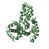
| Deposited unit | 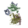
| ||||||||
|---|---|---|---|---|---|---|---|---|---|
| 1 | 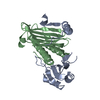
| ||||||||
| 2 | 
| ||||||||
| Unit cell |
| ||||||||
| Noncrystallographic symmetry (NCS) | NCS oper: (Code: given Matrix: (-0.02464, 0.98232, -0.18559), Vector: |
-Components
| #1: Protein | Mass: 19042.127 Da / Num. of mol.: 2 Source method: isolated from a genetically manipulated source Source: (gene. exp.)  #2: Protein | Mass: 17590.105 Da / Num. of mol.: 2 Source method: isolated from a genetically manipulated source Source: (gene. exp.) #3: Water | ChemComp-HOH / |  Mass: 18.015 Da / Num. of mol.: 100 / Source method: isolated from a natural source / Formula: H2O Mass: 18.015 Da / Num. of mol.: 100 / Source method: isolated from a natural source / Formula: H2O |
|---|
-Experimental details
-Experiment
| Experiment | Method: X-RAY DIFFRACTION / Number of used crystals: 1 |
|---|
- Sample preparation
| Crystal | Density Matthews: 2.3 Å3/Da / Density % sol: 55 % | ||||||||||||||||||
|---|---|---|---|---|---|---|---|---|---|---|---|---|---|---|---|---|---|---|---|
| Crystal grow | pH: 5.5 / Details: SODIUM CITRATE PH5.25 PEG 3000 12-20%, pH 5.50 | ||||||||||||||||||
| Crystal grow | *PLUS Temperature: 4 ℃ / Method: vapor diffusion | ||||||||||||||||||
| Components of the solutions | *PLUS
|
-Data collection
| Diffraction | Mean temperature: 100 K |
|---|---|
| Diffraction source | Source: SYNCHROTRON / Site: ESRF  / Beamline: ID14-1 / Wavelength: 0.933 / Beamline: ID14-1 / Wavelength: 0.933 |
| Detector | Type: ADSC CCD / Detector: CCD / Date: May 15, 2002 |
| Radiation | Protocol: SINGLE WAVELENGTH / Monochromatic (M) / Laue (L): M / Scattering type: x-ray |
| Radiation wavelength | Wavelength: 0.933 Å / Relative weight: 1 |
| Reflection | Resolution: 2.5→30 Å / Num. obs: 23227 / % possible obs: 99.4 % / Observed criterion σ(I): 2 / Redundancy: 4.1 % / Rmerge(I) obs: 0.05 / Net I/σ(I): 10.7 |
| Reflection shell | Resolution: 2.5→2.6 Å / Redundancy: 4.2 % / Rmerge(I) obs: 0.325 / Mean I/σ(I) obs: 2.3 / % possible all: 99.4 |
| Reflection | *PLUS Lowest resolution: 30 Å / Rmerge(I) obs: 0.055 |
| Reflection shell | *PLUS Lowest resolution: 2.64 Å / % possible obs: 99.4 % |
- Processing
| Software |
| ||||||||||||||||||||||||||||||||||||||||||||||||||||||||||||
|---|---|---|---|---|---|---|---|---|---|---|---|---|---|---|---|---|---|---|---|---|---|---|---|---|---|---|---|---|---|---|---|---|---|---|---|---|---|---|---|---|---|---|---|---|---|---|---|---|---|---|---|---|---|---|---|---|---|---|---|---|---|
| Refinement | Method to determine structure: MIR / Resolution: 2.5→30 Å / Cross valid method: THROUGHOUT / σ(F): 2
| ||||||||||||||||||||||||||||||||||||||||||||||||||||||||||||
| Displacement parameters | Biso mean: 31.3477 Å2 | ||||||||||||||||||||||||||||||||||||||||||||||||||||||||||||
| Refinement step | Cycle: LAST / Resolution: 2.5→30 Å
| ||||||||||||||||||||||||||||||||||||||||||||||||||||||||||||
| Refine LS restraints |
| ||||||||||||||||||||||||||||||||||||||||||||||||||||||||||||
| Refinement | *PLUS Lowest resolution: 30 Å | ||||||||||||||||||||||||||||||||||||||||||||||||||||||||||||
| Solvent computation | *PLUS | ||||||||||||||||||||||||||||||||||||||||||||||||||||||||||||
| Displacement parameters | *PLUS |
