- PDB-5w4w: Identification and Profiling of a Selective and Brain Penetrant R... -
+
Open data
ID or keywords:
Loading...
-
Basic information
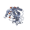
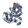
Entry
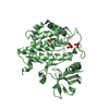
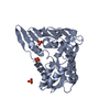
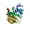
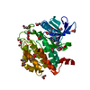
Database: PDB / ID: 5w4w
Title
Identification and Profiling of a Selective and Brain Penetrant Radioligand for In Vivo Target Occupancy Measurement of Casein Kinase 1 (CK1) Inhibitors
positive regulation of non-canonical Wnt signaling pathway / protein localization to Golgi apparatus / COPII vesicle coat assembly / protein localization to cilium / microtubule nucleation / tau-protein kinase / The CRY:PER:kinase complex represses transactivation by the BMAL:CLOCK (ARNTL:CLOCK) complex / midbrain dopaminergic neuron differentiation / non-motile cilium assembly / protein localization to centrosome ...positive regulation of non-canonical Wnt signaling pathway / protein localization to Golgi apparatus / COPII vesicle coat assembly / protein localization to cilium / microtubule nucleation / tau-protein kinase / The CRY:PER:kinase complex represses transactivation by the BMAL:CLOCK (ARNTL:CLOCK) complex / midbrain dopaminergic neuron differentiation / non-motile cilium assembly / protein localization to centrosome / COPII-mediated vesicle transport / Phosphorylation and nuclear translocation of the CRY:PER:kinase complex / tau-protein kinase activity / Golgi organization / Major pathway of rRNA processing in the nucleolus and cytosol / spindle assembly / Loss of Nlp from mitotic centrosomes / Loss of proteins required for interphase microtubule organization from the centrosome / endoplasmic reticulum-Golgi intermediate compartment membrane / Recruitment of mitotic centrosome proteins and complexes / Recruitment of NuMA to mitotic centrosomes / Anchoring of the basal body to the plasma membrane / AURKA Activation by TPX2 / spindle microtubule / circadian regulation of gene expression / regulation of circadian rhythm / Wnt signaling pathway / spindle / endocytosis / positive regulation of canonical Wnt signaling pathway / Regulation of PLK1 Activity at G2/M Transition / positive regulation of proteasomal ubiquitin-dependent protein catabolic process / ciliary basal body / protein phosphorylation / protein kinase activity / non-specific serine/threonine protein kinase / cadherin binding / protein serine kinase activity / protein serine/threonine kinase activity / centrosome / perinuclear region of cytoplasm / Golgi apparatus / signal transduction / nucleoplasm / ATP binding / nucleus / plasma membrane / cytosol / cytoplasm Similarity search - Function
: / Phosphorylase Kinase; domain 1 / Phosphorylase Kinase; domain 1 / Transferase(Phosphotransferase) domain 1 / Transferase(Phosphotransferase); domain 1 / Serine/threonine-protein kinase, active site / Serine/Threonine protein kinases active-site signature. / Protein kinase domain / Serine/Threonine protein kinases, catalytic domain / Protein kinase, ATP binding site ...: / Phosphorylase Kinase; domain 1 / Phosphorylase Kinase; domain 1 / Transferase(Phosphotransferase) domain 1 / Transferase(Phosphotransferase); domain 1 / Serine/threonine-protein kinase, active site / Serine/Threonine protein kinases active-site signature. / Protein kinase domain / Serine/Threonine protein kinases, catalytic domain / Protein kinase, ATP binding site / Protein kinases ATP-binding region signature. / Protein kinase domain profile. / Protein kinase domain / Protein kinase-like domain superfamily / 2-Layer Sandwich / Orthogonal Bundle / Mainly Alpha / Alpha Beta Similarity search - Domain/homology
In the structure databanks used in Yorodumi, some data are registered as the other names, "COVID-19 virus" and "2019-nCoV". Here are the details of the virus and the list of structure data.
Jan 31, 2019. EMDB accession codes are about to change! (news from PDBe EMDB page)
EMDB accession codes are about to change! (news from PDBe EMDB page)
The allocation of 4 digits for EMDB accession codes will soon come to an end. Whilst these codes will remain in use, new EMDB accession codes will include an additional digit and will expand incrementally as the available range of codes is exhausted. The current 4-digit format prefixed with “EMD-” (i.e. EMD-XXXX) will advance to a 5-digit format (i.e. EMD-XXXXX), and so on. It is currently estimated that the 4-digit codes will be depleted around Spring 2019, at which point the 5-digit format will come into force.
The EM Navigator/Yorodumi systems omit the EMD- prefix.
Related info.:Q: What is EMD? / ID/Accession-code notation in Yorodumi/EM Navigator
Yorodumi is a browser for structure data from EMDB, PDB, SASBDB, etc.
This page is also the successor to EM Navigator detail page, and also detail information page/front-end page for Omokage search.
The word "yorodu" (or yorozu) is an old Japanese word meaning "ten thousand". "mi" (miru) is to see.
Related info.:EMDB / PDB / SASBDB / Comparison of 3 databanks / Yorodumi Search / Aug 31, 2016. New EM Navigator & Yorodumi / Yorodumi Papers / Jmol/JSmol / Function and homology information / Changes in new EM Navigator and Yorodumi
 Movie
Movie Controller

 Yorodumi
 Open data
 Basic information
 Components
 Keywords
 Function and homology information
 Homo sapiens (human)
 X-RAY DIFFRACTION /
  Authors
 Citation
 Structure visualization
 Downloads & links
 Other downloads
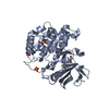

 PDBj
PDBj


 Assembly

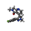
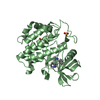
 Mass: 322.336 Da / Num. of mol.: 4 / Source method: obtained synthetically / Formula: C18H15FN4O
 Mass: 96.063 Da / Num. of mol.: 15 / Source method: obtained synthetically / Formula: SO4
 Mass: 18.015 Da / Num. of mol.: 519 / Source method: isolated from a natural source / Formula: H2O
 Sample preparation
 / Beamline: 17-ID / Wavelength: 1 Å
 Processing