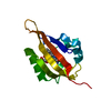
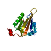
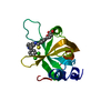
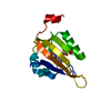
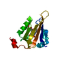
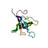
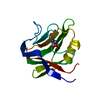
- PDB-6gpu: Crystal structure of miniSOG at 1.17A resolution -
+
Open data
ID or keywords:
Loading...
-
Basic information
Entry
Database: PDB / ID: 6gpu
Title
Crystal structure of miniSOG at 1.17A resolution
Components
Phototropin-2
Keywords
FLAVOPROTEIN / Singlet oxygen generator / fluorescent protein / FMN
Function / homology
Function and homology information
chloroplast relocation / phototropism / blue light signaling pathway / stomatal movement / blue light photoreceptor activity / response to blue light / plastid / circadian rhythm / kinase activity / FMN binding ...chloroplast relocation / phototropism / blue light signaling pathway / stomatal movement / blue light photoreceptor activity / response to blue light / plastid / circadian rhythm / kinase activity / FMN binding / non-specific serine/threonine protein kinase / protein serine kinase activity / protein serine/threonine kinase activity / Golgi apparatus / ATP binding / membrane / identical protein binding / nucleus / plasma membrane / cytoplasm Similarity search - Function
PAS domain / PAS-associated, C-terminal / PAC domain profile. / PAC motif / Motif C-terminal to PAS motifs (likely to contribute to PAS structural domain) / PAS domain / Beta-Lactamase / PAS domain / PAS repeat profile. / PAS domain ...PAS domain / PAS-associated, C-terminal / PAC domain profile. / PAC motif / Motif C-terminal to PAS motifs (likely to contribute to PAS structural domain) / PAS domain / Beta-Lactamase / PAS domain / PAS repeat profile. / PAS domain / PAS domain superfamily / Serine/threonine-protein kinase, active site / Serine/Threonine protein kinases active-site signature. / Protein kinase domain / Serine/Threonine protein kinases, catalytic domain / Protein kinase, ATP binding site / Protein kinases ATP-binding region signature. / Protein kinase domain profile. / Protein kinase domain / Protein kinase-like domain superfamily / 2-Layer Sandwich / Alpha Beta Similarity search - Domain/homology
Phototropin-2 / Defective in chloroplast avoidance protein 1 / Non-phototropic hypocotyl 1-like protein 1 / NPH1- ...Defective in chloroplast avoidance protein 1 / Non-phototropic hypocotyl 1-like protein 1 / NPH1-like protein 1
Mass: 13324.119 Da / Num. of mol.: 1 Source method: isolated from a genetically manipulated source Details: EFIPNPLLG / Source: (gene. exp.) Arabidopsis thaliana (thale cress) / Gene: PHOT2, CAV1, KIN7, NPL1, At5g58140, K21L19.6 / Production host: Escherichia coli BL21 (bacteria) References: UniProt: P93025, non-specific serine/threonine protein kinase
Resolution: 1.17→38.35 Å / Cor.coef. Fo:Fc: 0.981 / Cor.coef. Fo:Fc free: 0.972 / SU B: 1.289 / SU ML: 0.025 / Cross valid method: THROUGHOUT / ESU R: 0.036 / ESU R Free: 0.036 / Details: HYDROGENS HAVE BEEN ADDED IN THE RIDING POSITIONS
Rfactor
Num. reflection
% reflection
Selection details
Rfree
0.164
1850
4.9 %
RANDOM
Rwork
0.13265
-
-
-
obs
0.13414
36063
99.8 %
-
Solvent computation
Ion probe radii: 0.8 Å / Shrinkage radii: 0.8 Å / VDW probe radii: 1.2 Å
 Movie
Movie Controller


 Open data
 Basic information
 Components
 Keywords
 Function and homology information
 X-RAY DIFFRACTION /
  Authors
 France, 1items
  Citation
 Structure visualization
 Downloads & links
 Other downloads
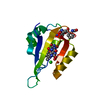


 PDBj
PDBj


 Assembly


 Mass: 456.344 Da / Num. of mol.: 1 / Source method: obtained synthetically / Formula: C17H21N4O9P
 Mass: 35.453 Da / Num. of mol.: 1 / Source method: obtained synthetically / Formula: Cl
 Mass: 24.305 Da / Num. of mol.: 1 / Source method: obtained synthetically / Formula: Mg
 Mass: 58.933 Da / Num. of mol.: 1 / Source method: obtained synthetically / Formula: Co
 Mass: 122.143 Da / Num. of mol.: 1 / Source method: obtained synthetically / Formula: C4H12NO3 / Comment: pH buffer*YM
 Sample preparation
 Processing