 Movie
Movie Controller


+ Open data

- Basic information
| Entry | Database: PDB / ID: 4aeh | ||||||
|---|---|---|---|---|---|---|---|
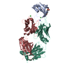
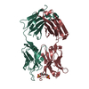
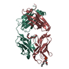
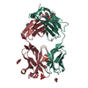
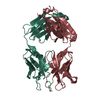
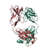
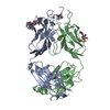
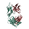
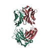
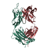
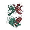
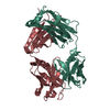
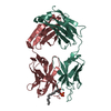
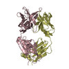
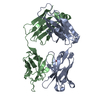
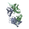
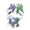
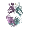
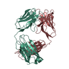
| Title | Crystal structure of the anti-AaHI Fab9C2 antibody | ||||||
 Components Components |
| ||||||
 Keywords Keywords | IMMUNE SYSTEM / ANDROCTONUS / ALPHA-TOXIN / COMBINING SITE / EPITOPE / PHARMACOLOGICAL SITE / VENOM / VOLTAGE- ACTIVATED SODIUM CHANNEL / SCORPION | ||||||
| Function / homology | Immunoglobulins / Immunoglobulin-like / Sandwich / Mainly Beta Function and homology information Function and homology information | ||||||
| Biological species |  | ||||||
| Method |  X-RAY DIFFRACTION / SYNCHROTRON / MOLECULAR REPLACEMENT / Resolution: 1.6 Å X-RAY DIFFRACTION / SYNCHROTRON / MOLECULAR REPLACEMENT / Resolution: 1.6 Å | ||||||
 Authors Authors | Fabrichny, I.P. / Mondielli, G. / Conrod, S. / Martin-Eauclaire, M.F. / Bourne, Y. / Marchot, P. | ||||||
 Citation Citation | Journal: J.Biol.Chem. / Year: 2012 Title: Structural Insights Into Antibody Sequestering and Neutralizing of Na+-Channel & [Alpha]-Type Modulator from Old-World Scorpion Venom Authors: Fabrichny, I.P. / Mondielli, G. / Conrod, S. / Martin-Eauclaire, M.F. / Bourne, Y. / Marchot, P. #1: Journal: Eur.J.Biochem. / Year: 2001 Title: Construction and Functional Evaluation of a Single-Chain Antibody Fragment that Neutralizes Toxin Aahi from the Venom of the Scorpion Androctonus Australis Hector. Authors: Devaux, C. / Moreau, E. / Goyffon, M. / Rochat, H. / Billiald, P. | ||||||
| History |
|
- Structure visualization
| Structure viewer | Molecule: MolmilJmol/JSmol |
|---|
- Downloads & links
-Download
| PDBx/mmCIF format | 4aeh.cif.gz | 196.3 KB | Display | PDBx/mmCIF format |
|---|---|---|---|---|
| PDB format | pdb4aeh.ent.gz | 157.9 KB | Display | PDB format |
| PDBx/mmJSON format | 4aeh.json.gz | Tree view | PDBx/mmJSON format | |
| Others |  Other downloads Other downloads |
-Validation report
| Arichive directory | https://data.pdbj.org/pub/pdb/validation_reports/ae/4aehftp://data.pdbj.org/pub/pdb/validation_reports/ae/4aeh | HTTPS FTP |
|---|
-Related structure data


-Links
 PDBj
PDBj

- Assembly
| Deposited unit | 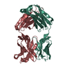
| ||||||||
|---|---|---|---|---|---|---|---|---|---|
| 1 |
| ||||||||
| Unit cell |
|
-Components
| #1: Antibody | Mass: 24524.275 Da / Num. of mol.: 1 / Fragment: VARIABLE DOMAIN, RESIDUES 1-231 / Source method: isolated from a natural source / Details: IGG2A, K-ISSUED ANTIBODY / Source: (natural) |
|---|---|
| #2: Antibody | Mass: 23678.111 Da / Num. of mol.: 1 / Fragment: VARIABLE DOMAIN, RESIDUES 1-214 / Source method: isolated from a natural source / Details: IGG2A, K-ISSUED ANTIBODY / Source: (natural) |
| #3: Water | ChemComp-HOH /  Mass: 18.015 Da / Num. of mol.: 614 / Source method: isolated from a natural source / Formula: H2O Mass: 18.015 Da / Num. of mol.: 614 / Source method: isolated from a natural source / Formula: H2O |
| Has protein modification | Y |
| Sequence details | SEE REFERENCE IN REMARK 1 FOR SEQUENCES OF CHAIN L AND CHAIN H. |
-Experimental details
-Experiment
| Experiment | Method: X-RAY DIFFRACTION / Number of used crystals: 1 |
|---|
- Sample preparation
| Crystal | Density Matthews: 2.5 Å3/Da / Density % sol: 51 % / Description: NONE |
|---|---|
| Crystal grow | pH: 7 Details: 12.5% PEG 4000, 0.1 M IMIDAZOLE-MALATE PH 7.0, 50 MM NACL |
-Data collection
| Diffraction | Mean temperature: 100 K |
|---|---|
| Diffraction source | Source: SYNCHROTRON / Site: ESRF  / Beamline: ID23-1 / Wavelength: 0.9798 / Beamline: ID23-1 / Wavelength: 0.9798 |
| Detector | Type: ADSC QUANTUM 315r / Detector: CCD / Date: Dec 10, 2007 |
| Radiation | Protocol: SINGLE WAVELENGTH / Monochromatic (M) / Laue (L): M / Scattering type: x-ray |
| Radiation wavelength | Wavelength: 0.9798 Å / Relative weight: 1 |
| Reflection | Resolution: 1.6→15 Å / Num. obs: 63057 / % possible obs: 99.5 % / Observed criterion σ(I): 2 / Redundancy: 3.7 % / Biso Wilson estimate: 37.4 Å2 / Rmerge(I) obs: 0.06 / Net I/σ(I): 11.26 |
| Reflection shell | Resolution: 1.6→1.65 Å / Redundancy: 3.7 % / Rmerge(I) obs: 0.42 / Mean I/σ(I) obs: 2.8 / % possible all: 99.7 |
- Processing
| Software |
| ||||||||||||||||||||||||||||||||||||||||||||||||||||||||||||||||||||||||||||||||||||||||||||||||||||||||||||||||||||||||||||||||||||||||||||||||||||||||||||||||||||||||||||||||||||||
|---|---|---|---|---|---|---|---|---|---|---|---|---|---|---|---|---|---|---|---|---|---|---|---|---|---|---|---|---|---|---|---|---|---|---|---|---|---|---|---|---|---|---|---|---|---|---|---|---|---|---|---|---|---|---|---|---|---|---|---|---|---|---|---|---|---|---|---|---|---|---|---|---|---|---|---|---|---|---|---|---|---|---|---|---|---|---|---|---|---|---|---|---|---|---|---|---|---|---|---|---|---|---|---|---|---|---|---|---|---|---|---|---|---|---|---|---|---|---|---|---|---|---|---|---|---|---|---|---|---|---|---|---|---|---|---|---|---|---|---|---|---|---|---|---|---|---|---|---|---|---|---|---|---|---|---|---|---|---|---|---|---|---|---|---|---|---|---|---|---|---|---|---|---|---|---|---|---|---|---|---|---|---|---|
| Refinement | Method to determine structure: MOLECULAR REPLACEMENT / Resolution: 1.6→30 Å / Cor.coef. Fo:Fc: 0.966 / Cor.coef. Fo:Fc free: 0.95 / SU B: 3.777 / SU ML: 0.066 / Cross valid method: THROUGHOUT / ESU R: 0.088 / ESU R Free: 0.092 / Stereochemistry target values: MAXIMUM LIKELIHOOD Details: HYDROGENS HAVE BEEN ADDED IN THE RIDING POSITIONS. U VALUES WITH TLS ADDED. HYDROGENS HAVE BEEN USED IF PRESENT IN THE INPUT.
| ||||||||||||||||||||||||||||||||||||||||||||||||||||||||||||||||||||||||||||||||||||||||||||||||||||||||||||||||||||||||||||||||||||||||||||||||||||||||||||||||||||||||||||||||||||||
| Solvent computation | Ion probe radii: 0.8 Å / Shrinkage radii: 0.8 Å / VDW probe radii: 1.2 Å / Solvent model: BABINET MODEL WITH MASK | ||||||||||||||||||||||||||||||||||||||||||||||||||||||||||||||||||||||||||||||||||||||||||||||||||||||||||||||||||||||||||||||||||||||||||||||||||||||||||||||||||||||||||||||||||||||
| Displacement parameters | Biso mean: 22.612 Å2
| ||||||||||||||||||||||||||||||||||||||||||||||||||||||||||||||||||||||||||||||||||||||||||||||||||||||||||||||||||||||||||||||||||||||||||||||||||||||||||||||||||||||||||||||||||||||
| Refinement step | Cycle: LAST / Resolution: 1.6→30 Å
| ||||||||||||||||||||||||||||||||||||||||||||||||||||||||||||||||||||||||||||||||||||||||||||||||||||||||||||||||||||||||||||||||||||||||||||||||||||||||||||||||||||||||||||||||||||||
| Refine LS restraints |
|