SHEET DETERMINATION METHOD: DSSP THE SHEETS PRESENTED AS "BA" IN EACH CHAIN ON SHEET RECORDS BELOW ... SHEET DETERMINATION METHOD: DSSP THE SHEETS PRESENTED AS "BA" IN EACH CHAIN ON SHEET RECORDS BELOW IS ACTUALLY AN 5-STRANDED BARREL THIS IS REPRESENTED BY A 6-STRANDED SHEET IN WHICH THE FIRST AND LAST STRANDS ARE IDENTICAL.
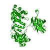
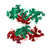
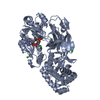
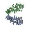
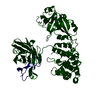
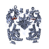
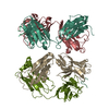
DIPHTHERIATOXIN / DT / NAD(+)--DIPHTHAMIDE ADP-RIBOSYLTRANSFERASE / DIPHTHERIA TOXIN FRAGMENT A / DIPHTHERIA TOXIN ...DT / NAD(+)--DIPHTHAMIDE ADP-RIBOSYLTRANSFERASE / DIPHTHERIA TOXIN FRAGMENT A / DIPHTHERIA TOXIN FRAGMENT B / CRM197
Mass: 58483.422 Da / Num. of mol.: 2 / Mutation: YES / Source method: isolated from a natural source / Source: (natural) CORYNEBACTERIUM DIPHTHERIAE (bacteria) References: UniProt: P00588, NAD+-diphthamide ADP-ribosyltransferase
Mass: 18.015 Da / Num. of mol.: 252 / Source method: isolated from a natural source / Formula: H2O
Compound details
ENGINEERED RESIDUE IN CHAIN A, GLY 53 TO GLU ENGINEERED RESIDUE IN CHAIN B, GLY 53 TO GLU
Has protein modification
Y
Sequence details
COORDINATES USE STANDARD RESIDUE NUMBERING FOR CONSISTENCY WITH PREVIOUS WORK RESULTING IN THE ...COORDINATES USE STANDARD RESIDUE NUMBERING FOR CONSISTENCY WITH PREVIOUS WORK RESULTING IN THE NATURAL GLY TO GLU MUTATION AT POSITION 52 INSTEAD OF 53. RESIDUES 1-32 IN THE UNIPROT SEQUENCE ARE THE SIGNAL PEPTIDE.
-
Experimental details
-
Experiment
Experiment
Method: X-RAY DIFFRACTION / Number of used crystals: 1
-
Sample preparation
Crystal
Density Matthews: 2.87 Å3/Da / Density % sol: 57 % / Description: NONE
Crystal grow
pH: 9 / Details: 1.9 M AMMONIUM SULFATE, 100 MM BICINE [PH 9.0]
Resolution: 2.078→52.561 Å / SU ML: 0.34 / σ(F): 0.01 / Phase error: 26.05 / Stereochemistry target values: ML Details: RESIDUES OF CHAIN A 38-50, 188-199, 350-353, 502-503, AND 517-519, ARE DISORDERED. AND RESIDUES OF CHAIN B 1-3, 38-50, 188-200, 352-353 AND 517-520, ARE DISORDERED. DISORDERED SIDE CHAINS ...Details: RESIDUES OF CHAIN A 38-50, 188-199, 350-353, 502-503, AND 517-519, ARE DISORDERED. AND RESIDUES OF CHAIN B 1-3, 38-50, 188-200, 352-353 AND 517-520, ARE DISORDERED. DISORDERED SIDE CHAINS WERE MODELED UP TO THE BACKBONE BETA ATOMS, AND RESIDUE NAMES WERE KEPT CONSISTENT WITH WITH THE SEQUENCE OF THE PROTEIN.
Rfactor
Num. reflection
% reflection
Rfree
0.2439
3019
5.1 %
Rwork
0.1955
-
-
obs
0.198
59591
83.68 %
Solvent computation
Shrinkage radii: 0.9 Å / VDW probe radii: 1.11 Å / Solvent model: FLAT BULK SOLVENT MODEL / Bsol: 41.156 Å2 / ksol: 0.367 e/Å3
Displacement parameters
Biso mean: 40 Å2
Baniso -1
Baniso -2
Baniso -3
1-
-1.7665 Å2
0.1379 Å2
0.4956 Å2
2-
-
4.0574 Å2
-0.1731 Å2
3-
-
-
-2.2909 Å2
Refinement step
Cycle: LAST / Resolution: 2.078→52.561 Å
Protein
Nucleic acid
Ligand
Solvent
Total
Num. atoms
7446
0
18
252
7716
Refine LS restraints
Refine-ID
Type
Dev ideal
Number
X-RAY DIFFRACTION
f_bond_d
0.007
7627
X-RAY DIFFRACTION
f_angle_d
1.059
10371
X-RAY DIFFRACTION
f_dihedral_angle_d
13.57
2657
X-RAY DIFFRACTION
f_chiral_restr
0.071
1198
X-RAY DIFFRACTION
f_plane_restr
0.005
1356
LS refinement shell
Resolution (Å)
Rfactor Rfree
Num. reflection Rfree
Rfactor Rwork
Num. reflection Rwork
Refine-ID
% reflection obs (%)
2.078-2.1523
0.3281
266
0.2629
4630
X-RAY DIFFRACTION
68
2.1523-2.2385
0.3177
247
0.2556
4829
X-RAY DIFFRACTION
71
2.2385-2.3404
0.3172
282
0.2331
5188
X-RAY DIFFRACTION
77
2.3404-2.4637
0.264
264
0.2124
5513
X-RAY DIFFRACTION
81
2.4637-2.6181
0.2802
307
0.2009
5743
X-RAY DIFFRACTION
85
2.6181-2.8202
0.2356
320
0.1934
5912
X-RAY DIFFRACTION
88
2.8202-3.104
0.2372
331
0.1918
6081
X-RAY DIFFRACTION
90
3.104-3.5531
0.2326
348
0.1836
6162
X-RAY DIFFRACTION
92
3.5531-4.4761
0.2108
328
0.1569
6214
X-RAY DIFFRACTION
92
4.4761-52.5777
0.2154
326
0.1864
6300
X-RAY DIFFRACTION
93
Refinement TLS params.
Method: refined / Origin x: 1.349 Å / Origin y: -48.4499 Å / Origin z: 30.4154 Å
11
12
13
21
22
23
31
32
33
T
0.0964 Å2
0.001 Å2
0.0214 Å2
-
0.0594 Å2
-0.0067 Å2
-
-
0.0447 Å2
L
0.9647 °2
-0.2986 °2
0.1738 °2
-
0.6633 °2
-0.0917 °2
-
-
0.6882 °2
S
0.0983 Å °
0.0712 Å °
-0.026 Å °
-0.0537 Å °
-0.0818 Å °
0.0734 Å °
0.027 Å °
0.0876 Å °
-0.0171 Å °
Refinement TLS group
Selection details: ALL
+
About Yorodumi
-
News
-
Feb 9, 2022. New format data for meta-information of EMDB entries
New format data for meta-information of EMDB entries
Version 3 of the EMDB header file is now the official format.
The previous official version 1.9 will be removed from the archive.
In the structure databanks used in Yorodumi, some data are registered as the other names, "COVID-19 virus" and "2019-nCoV". Here are the details of the virus and the list of structure data.
Jan 31, 2019. EMDB accession codes are about to change! (news from PDBe EMDB page)
EMDB accession codes are about to change! (news from PDBe EMDB page)
The allocation of 4 digits for EMDB accession codes will soon come to an end. Whilst these codes will remain in use, new EMDB accession codes will include an additional digit and will expand incrementally as the available range of codes is exhausted. The current 4-digit format prefixed with “EMD-” (i.e. EMD-XXXX) will advance to a 5-digit format (i.e. EMD-XXXXX), and so on. It is currently estimated that the 4-digit codes will be depleted around Spring 2019, at which point the 5-digit format will come into force.
The EM Navigator/Yorodumi systems omit the EMD- prefix.
Related info.:Q: What is EMD? / ID/Accession-code notation in Yorodumi/EM Navigator
Yorodumi is a browser for structure data from EMDB, PDB, SASBDB, etc.
This page is also the successor to EM Navigator detail page, and also detail information page/front-end page for Omokage search.
The word "yorodu" (or yorozu) is an old Japanese word meaning "ten thousand". "mi" (miru) is to see.
Related info.:EMDB / PDB / SASBDB / Comparison of 3 databanks / Yorodumi Search / Aug 31, 2016. New EM Navigator & Yorodumi / Yorodumi Papers / Jmol/JSmol / Function and homology information / Changes in new EM Navigator and Yorodumi
 Movie
Movie Controller

 Yorodumi
 Open data
 Basic information
 Components
 Keywords
 Function and homology information
 CORYNEBACTERIUM DIPHTHERIAE (bacteria)
 X-RAY DIFFRACTION /
  Authors
 Citation
 Structure visualization
 Downloads & links
 Other downloads
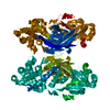


 PDBj
PDBj
 Assembly
 Mass: 122.125 Da / Num. of mol.: 2 / Source method: obtained synthetically / Formula: C6H6N2O / Comment: medication*YM
 Mass: 18.015 Da / Num. of mol.: 252 / Source method: isolated from a natural source / Formula: H2O
 Sample preparation
 / Beamline: 5.0.3 / Wavelength: 0.979
  Processing