 Movie
Movie Controller

[English] 日本語
 Yorodumi
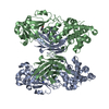
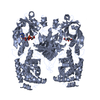
- PDB-7k7e: Crystal structure of diphtheria toxin from crystals obtained at pH 7.0 -
+ Open data

- Basic information
| Entry | Database: PDB / ID: 7k7e | |||||||||
|---|---|---|---|---|---|---|---|---|---|---|
| Title | Crystal structure of diphtheria toxin from crystals obtained at pH 7.0 | |||||||||
 Components Components | Diphtheria toxin | |||||||||
 Keywords Keywords | TOXIN / diptheria toxin / pH dependent conformational changes | |||||||||
| Function / homology |  Function and homology information Function and homology informationNAD+-diphthamide ADP-ribosyltransferase / NAD+-diphthamide ADP-ribosyltransferase activity / toxin activity / : / identical protein binding Similarity search - Function | |||||||||
| Biological species |  Corynebacterium diphtheriae (bacteria) Corynebacterium diphtheriae (bacteria) | |||||||||
| Method |  X-RAY DIFFRACTION / SYNCHROTRON / MOLECULAR REPLACEMENT / molecular replacement / Resolution: 2.3 Å X-RAY DIFFRACTION / SYNCHROTRON / MOLECULAR REPLACEMENT / molecular replacement / Resolution: 2.3 Å | |||||||||
 Authors Authors | Lovell, S. / Kashipathy, M.M. / Battaile, K.P. / Rodnin, M.V. / Ladokhin, A.S. | |||||||||
| Funding support |  United States, 2items United States, 2items
| |||||||||
 Citation Citation | Journal: Toxins / Year: 2020 Title: Structure of the Diphtheria Toxin at Acidic pH: Implications for the Conformational Switching of the Translocation Domain. Authors: Rodnin, M.V. / Kashipathy, M.M. / Kyrychenko, A. / Battaile, K.P. / Lovell, S. / Ladokhin, A.S. | |||||||||
| History |
|
- Structure visualization
| Structure viewer | Molecule: MolmilJmol/JSmol |
|---|
- Downloads & links
-Download
| PDBx/mmCIF format | 7k7e.cif.gz | 206.1 KB | Display | PDBx/mmCIF format |
|---|---|---|---|---|
| PDB format | pdb7k7e.ent.gz | 161.9 KB | Display | PDB format |
| PDBx/mmJSON format | 7k7e.json.gz | Tree view | PDBx/mmJSON format | |
| Others |  Other downloads Other downloads |
-Validation report
| Arichive directory | https://data.pdbj.org/pub/pdb/validation_reports/k7/7k7eftp://data.pdbj.org/pub/pdb/validation_reports/k7/7k7e | HTTPS FTP |
|---|
-Related structure data
| Related structure data |  7k7bC 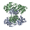 7k7cC 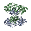 7k7dC 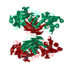 1ddtS S: Starting model for refinement C: citing same article ( |
|---|---|
| Similar structure data |


-Links
 PDBj
PDBj
- Assembly
| Deposited unit | 
| ||||||||
|---|---|---|---|---|---|---|---|---|---|
| 1 |
| ||||||||
| Unit cell |
|
-Components
| #1: Protein | Mass: 58744.828 Da / Num. of mol.: 2 / Fragment: Full Length / Mutation: K51E, E148K Source method: isolated from a genetically manipulated source Source: (gene. exp.) Corynebacterium diphtheriae (bacteria) / Plasmid: pET15b / Production host: #2: Water | ChemComp-HOH / |  Mass: 18.015 Da / Num. of mol.: 201 / Source method: isolated from a natural source / Formula: H2O Mass: 18.015 Da / Num. of mol.: 201 / Source method: isolated from a natural source / Formula: H2OHas protein modification | Y | |
|---|
-Experimental details
-Experiment
| Experiment | Method: X-RAY DIFFRACTION / Number of used crystals: 1 |
|---|
- Sample preparation
| Crystal | Density Matthews: 2.61 Å3/Da / Density % sol: 52.96 % / Mosaicity: 0.25 ° |
|---|---|
| Crystal grow | Temperature: 291 K / Method: vapor diffusion, sitting drop / pH: 7 Details: 10% (w/v) PEG 10000, 0.1 M magnesium acetate, 0.1 M HEPES |
-Data collection
| Diffraction | Mean temperature: 100 K / Serial crystal experiment: N | ||||||||||||||||||||||||
|---|---|---|---|---|---|---|---|---|---|---|---|---|---|---|---|---|---|---|---|---|---|---|---|---|---|
| Diffraction source | Source: SYNCHROTRON / Site: APS / Beamline: 17-ID / Wavelength: 1 Å | ||||||||||||||||||||||||
| Detector | Type: DECTRIS PILATUS 6M / Detector: PIXEL / Date: Apr 15, 2018 | ||||||||||||||||||||||||
| Radiation | Protocol: SINGLE WAVELENGTH / Monochromatic (M) / Laue (L): M / Scattering type: x-ray | ||||||||||||||||||||||||
| Radiation wavelength | Wavelength: 1 Å / Relative weight: 1 | ||||||||||||||||||||||||
| Reflection | Resolution: 2.3→46.74 Å / Num. obs: 51695 / % possible obs: 97.7 % / Redundancy: 3.5 % / CC1/2: 0.997 / Rmerge(I) obs: 0.073 / Net I/σ(I): 9.7 / Num. measured all: 183332 | ||||||||||||||||||||||||
| Reflection shell | Diffraction-ID: 1
|
-Phasing
| Phasing | Method: molecular replacement |
|---|
- Processing
| Software |
| ||||||||||||||||||||||||||||||||||||||||||||||||||||||||||||||||||||||||||||||||||||||||||||||||||||||||||||
|---|---|---|---|---|---|---|---|---|---|---|---|---|---|---|---|---|---|---|---|---|---|---|---|---|---|---|---|---|---|---|---|---|---|---|---|---|---|---|---|---|---|---|---|---|---|---|---|---|---|---|---|---|---|---|---|---|---|---|---|---|---|---|---|---|---|---|---|---|---|---|---|---|---|---|---|---|---|---|---|---|---|---|---|---|---|---|---|---|---|---|---|---|---|---|---|---|---|---|---|---|---|---|---|---|---|---|---|---|---|
| Refinement | Method to determine structure: MOLECULAR REPLACEMENT Starting model: 1DDT Resolution: 2.3→32.721 Å / SU ML: 0.32 / Cross valid method: THROUGHOUT / σ(F): 1.05 / Phase error: 30.11 / Stereochemistry target values: ML
| ||||||||||||||||||||||||||||||||||||||||||||||||||||||||||||||||||||||||||||||||||||||||||||||||||||||||||||
| Solvent computation | Shrinkage radii: 0.9 Å / VDW probe radii: 1.11 Å / Solvent model: FLAT BULK SOLVENT MODEL | ||||||||||||||||||||||||||||||||||||||||||||||||||||||||||||||||||||||||||||||||||||||||||||||||||||||||||||
| Displacement parameters | Biso max: 120.67 Å2 / Biso mean: 56.3183 Å2 / Biso min: 24.31 Å2 | ||||||||||||||||||||||||||||||||||||||||||||||||||||||||||||||||||||||||||||||||||||||||||||||||||||||||||||
| Refinement step | Cycle: final / Resolution: 2.3→32.721 Å
| ||||||||||||||||||||||||||||||||||||||||||||||||||||||||||||||||||||||||||||||||||||||||||||||||||||||||||||
| Refine LS restraints |
| ||||||||||||||||||||||||||||||||||||||||||||||||||||||||||||||||||||||||||||||||||||||||||||||||||||||||||||
| LS refinement shell | Refine-ID: X-RAY DIFFRACTION / Rfactor Rfree error: 0
|
