 Movie
Movie Controller


+ Open data

- Basic information
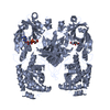
| Entry | Database: PDB / ID: 1sgk | ||||||
|---|---|---|---|---|---|---|---|
| Title | NUCLEOTIDE-FREE DIPHTHERIA TOXIN | ||||||
 Components Components | DIPHTHERIA TOXIN (DIMERIC) | ||||||
 Keywords Keywords | TOXIN / ADP-RIBOSYLATION / TRANSFERASE / GLYCOSYLTRANSFERASE / NAD / ADP-RIBOSYL TRANSFERASE | ||||||
| Function / homology |  Function and homology information Function and homology informationNAD+-diphthamide ADP-ribosyltransferase / NAD+-diphthamide ADP-ribosyltransferase activity / Uptake and function of diphtheria toxin / transmembrane protein transporter activity / nucleotidyltransferase activity / toxin activity / : / extracellular region / identical protein binding / plasma membrane Similarity search - Function | ||||||
| Biological species |  Corynephage beta (virus) Corynephage beta (virus) | ||||||
| Method |  X-RAY DIFFRACTION / MOLECULAR REPLACEMENT / Resolution: 2.3 Å X-RAY DIFFRACTION / MOLECULAR REPLACEMENT / Resolution: 2.3 Å | ||||||
 Authors Authors | Bell, C.E. / Eisenberg, D. | ||||||
 Citation Citation | Journal: Biochemistry / Year: 1997 Title: Crystal structure of nucleotide-free diphtheria toxin. Authors: Bell, C.E. / Eisenberg, D. #1: Journal: Biochemistry / Year: 1996Title: Crystal Structure of Diphtheria Toxin Bound to Nicotinamide Adenine Dinucleotide Authors: Bell, C.E. / Eisenberg, D. #2: Journal: Proc.Natl.Acad.Sci.USA / Year: 1994Title: Domain Swapping: Entangling Alliances between Proteins Authors: Bennett, M.J. / Choe, S. / Eisenberg, D. #3: Journal: Protein Sci. / Year: 1994Title: Refined Structure of Monomeric Diphtheria Toxin at 2.3 A Resolution Authors: Bennett, M.J. / Eisenberg, D. #4: Journal: Protein Sci. / Year: 1994Title: Refined Structure of Dimeric Diphtheria Toxin at 2.0 A Resolution Authors: Bennett, M.J. / Choe, S. / Eisenberg, D. #5: Journal: Nature / Year: 1992Title: The Crystal Structure of Diphtheria Toxin Authors: Choe, S. / Bennett, M.J. / Fujii, G. / Curmi, P.M. / Kantardjieff, K.A. / Collier, R.J. / Eisenberg, D. | ||||||
| History |
|
- Structure visualization
| Structure viewer | Molecule: MolmilJmol/JSmol |
|---|
- Downloads & links
-Download
| PDBx/mmCIF format | 1sgk.cif.gz | 120 KB | Display | PDBx/mmCIF format |
|---|---|---|---|---|
| PDB format | pdb1sgk.ent.gz | 92.4 KB | Display | PDB format |
| PDBx/mmJSON format | 1sgk.json.gz | Tree view | PDBx/mmJSON format | |
| Others |  Other downloads Other downloads |
-Validation report
| Arichive directory | https://data.pdbj.org/pub/pdb/validation_reports/sg/1sgkftp://data.pdbj.org/pub/pdb/validation_reports/sg/1sgk | HTTPS FTP |
|---|
-Related structure data
| Related structure data | 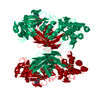 1ddtS S: Starting model for refinement |
|---|---|
| Similar structure data |


-Links
 PDBj
PDBj
- Assembly
| Deposited unit | 
| ||||||||
|---|---|---|---|---|---|---|---|---|---|
| 1 | 
| ||||||||
| Unit cell |
|
-Components
| #1: Protein | Mass: 58411.359 Da / Num. of mol.: 1 / Source method: isolated from a natural source / Details: PURCHASED FROM CONNAUGHT LABORATORIES / Source: (natural) Corynephage beta (virus) / Genus: Lambda-like virusesReferences: UniProt: P00588, NAD+-diphthamide ADP-ribosyltransferase |
|---|---|
| #2: Water | ChemComp-HOH /  Mass: 18.015 Da / Num. of mol.: 357 / Source method: isolated from a natural source / Formula: H2O Mass: 18.015 Da / Num. of mol.: 357 / Source method: isolated from a natural source / Formula: H2O |
| Has protein modification | Y |
-Experimental details
-Experiment
| Experiment | Method: X-RAY DIFFRACTION / Number of used crystals: 1 |
|---|
- Sample preparation
| Crystal | Density Matthews: 2.67 Å3/Da / Density % sol: 46 % | ||||||||||||||||||||||||||||||||||||||||||||||||
|---|---|---|---|---|---|---|---|---|---|---|---|---|---|---|---|---|---|---|---|---|---|---|---|---|---|---|---|---|---|---|---|---|---|---|---|---|---|---|---|---|---|---|---|---|---|---|---|---|---|
| Crystal grow | Method: vapor diffusion, hanging drop / pH: 7.5 Details: HANGING DROP, 12 % PEG8000, 0.43M NACL, 0.043M TRIS, PH 7.5, vapor diffusion - hanging drop | ||||||||||||||||||||||||||||||||||||||||||||||||
| Crystal | *PLUS | ||||||||||||||||||||||||||||||||||||||||||||||||
| Crystal grow | *PLUS Method: vapor diffusion, hanging drop | ||||||||||||||||||||||||||||||||||||||||||||||||
| Components of the solutions | *PLUS
|
-Data collection
| Diffraction | Mean temperature: 94 K |
|---|---|
| Diffraction source | Wavelength: 1.5418 |
| Detector | Type: RIGAKU / Detector: IMAGE PLATE / Date: Apr 15, 1994 |
| Radiation | Monochromatic (M) / Laue (L): M / Scattering type: x-ray |
| Radiation wavelength | Wavelength: 1.5418 Å / Relative weight: 1 |
| Reflection | Highest resolution: 2.2 Å / Num. obs: 26627 / % possible obs: 95.7 % / Observed criterion σ(I): 0 / Redundancy: 2.8 % / Biso Wilson estimate: 33.9 Å2 / Rmerge(I) obs: 0.066 / Net I/σ(I): 9.6 |
| Reflection shell | Resolution: 2.3→2.4 Å / Rmerge(I) obs: 0.239 / Mean I/σ(I) obs: 2.6 / % possible all: 92.7 |
| Reflection | *PLUS Highest resolution: 2.3 Å / Num. measured all: 74281 |
| Reflection shell | *PLUS % possible obs: 92.7 % |
- Processing
| Software |
| ||||||||||||||||||||||||||||||||||||||||||||||||||||||||||||||||||||||||||||||||
|---|---|---|---|---|---|---|---|---|---|---|---|---|---|---|---|---|---|---|---|---|---|---|---|---|---|---|---|---|---|---|---|---|---|---|---|---|---|---|---|---|---|---|---|---|---|---|---|---|---|---|---|---|---|---|---|---|---|---|---|---|---|---|---|---|---|---|---|---|---|---|---|---|---|---|---|---|---|---|---|---|---|
| Refinement | Method to determine structure: MOLECULAR REPLACEMENT Starting model: DT-DIMER APUP COMPLEX (PDB ENTRY 1DDT) Resolution: 2.3→8 Å / Cross valid method: FREE R-FACTOR / σ(F): 0
| ||||||||||||||||||||||||||||||||||||||||||||||||||||||||||||||||||||||||||||||||
| Displacement parameters | Biso mean: 38.8 Å2 | ||||||||||||||||||||||||||||||||||||||||||||||||||||||||||||||||||||||||||||||||
| Refine analyze |
| ||||||||||||||||||||||||||||||||||||||||||||||||||||||||||||||||||||||||||||||||
| Refinement step | Cycle: LAST / Resolution: 2.3→8 Å
| ||||||||||||||||||||||||||||||||||||||||||||||||||||||||||||||||||||||||||||||||
| Refine LS restraints |
| ||||||||||||||||||||||||||||||||||||||||||||||||||||||||||||||||||||||||||||||||
| LS refinement shell | Resolution: 2.3→2.4 Å
| ||||||||||||||||||||||||||||||||||||||||||||||||||||||||||||||||||||||||||||||||
| Xplor file |
| ||||||||||||||||||||||||||||||||||||||||||||||||||||||||||||||||||||||||||||||||
| Software | *PLUS Name: X-PLOR / Classification: refinement | ||||||||||||||||||||||||||||||||||||||||||||||||||||||||||||||||||||||||||||||||
| Refinement | *PLUS Num. reflection obs: 23161 | ||||||||||||||||||||||||||||||||||||||||||||||||||||||||||||||||||||||||||||||||
| Solvent computation | *PLUS | ||||||||||||||||||||||||||||||||||||||||||||||||||||||||||||||||||||||||||||||||
| Displacement parameters | *PLUS |
