 Movie
Movie Controller

[English] 日本語
 Yorodumi
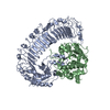
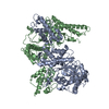
- PDB-1mdt: THE REFINED STRUCTURE OF MONOMERIC DIPHTHERIA TOXIN AT 2.3 ANGSTR... -
+ Open data

- Basic information
| Entry | Database: PDB / ID: 1mdt | ||||||
|---|---|---|---|---|---|---|---|
| Title | THE REFINED STRUCTURE OF MONOMERIC DIPHTHERIA TOXIN AT 2.3 ANGSTROMS RESOLUTION | ||||||
 Components Components | DIPHTHERIA TOXIN | ||||||
 Keywords Keywords | TOXIN | ||||||
| Function / homology |  Function and homology information Function and homology informationNAD+-diphthamide ADP-ribosyltransferase / NAD+-diphthamide ADP-ribosyltransferase activity / Uptake and function of diphtheria toxin / transmembrane protein transporter activity / nucleotidyltransferase activity / toxin activity / : / extracellular region / identical protein binding / plasma membrane Similarity search - Function | ||||||
| Biological species |  Corynephage beta (virus) Corynephage beta (virus) | ||||||
| Method |  X-RAY DIFFRACTION / Resolution: 2.3 Å X-RAY DIFFRACTION / Resolution: 2.3 Å | ||||||
 Authors Authors | Bennett, M.J. / Eisenberg, D. | ||||||
 Citation Citation | Journal: Protein Sci. / Year: 1994 Title: Refined structure of monomeric diphtheria toxin at 2.3 A resolution. Authors: Bennett, M.J. / Eisenberg, D. #1: Journal: Proc.Natl.Acad.Sci.USA / Year: 1994Title: Domain Swapping: Entangling Alliances between Proteins Authors: Bennett, M.J. / Choe, S. / Eisenberg, D. | ||||||
| History |
| ||||||
| Remark 700 | SHEET SHEET R1 IS NOT A CLOSED BARREL, BUT IT IS CLOSED ON ONE SIDE AND FLATTENED. |
- Structure visualization
| Structure viewer | Molecule: MolmilJmol/JSmol |
|---|
- Downloads & links
-Download
| PDBx/mmCIF format | 1mdt.cif.gz | 212.6 KB | Display | PDBx/mmCIF format |
|---|---|---|---|---|
| PDB format | pdb1mdt.ent.gz | 169.9 KB | Display | PDB format |
| PDBx/mmJSON format | 1mdt.json.gz | Tree view | PDBx/mmJSON format | |
| Others |  Other downloads Other downloads |
-Validation report
| Arichive directory | https://data.pdbj.org/pub/pdb/validation_reports/md/1mdtftp://data.pdbj.org/pub/pdb/validation_reports/md/1mdt | HTTPS FTP |
|---|
-Related structure data
| Similar structure data |
|---|


-Links
 PDBj
PDBj
- Assembly
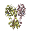
| Deposited unit | 
| ||||||||
|---|---|---|---|---|---|---|---|---|---|
| 1 |
| ||||||||
| Unit cell |
| ||||||||
| Noncrystallographic symmetry (NCS) | NCS oper: (Code: given Matrix: (-0.9995, -0.0296, -0.0078), Vector: |
-Components
| #1: Protein | Mass: 58411.359 Da / Num. of mol.: 2 Source method: isolated from a genetically manipulated source Source: (gene. exp.) Corynephage beta (virus) / Genus: Lambda-like viruses / References: UniProt: P00588#2: Chemical | 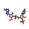  Mass: 653.387 Da / Num. of mol.: 2 / Source method: obtained synthetically / Formula: C19H25N7O15P2 Mass: 653.387 Da / Num. of mol.: 2 / Source method: obtained synthetically / Formula: C19H25N7O15P2#3: Water | ChemComp-HOH / |  Mass: 18.015 Da / Num. of mol.: 396 / Source method: isolated from a natural source / Formula: H2O Mass: 18.015 Da / Num. of mol.: 396 / Source method: isolated from a natural source / Formula: H2OHas protein modification | Y | |
|---|
-Experimental details
-Experiment
| Experiment | Method: X-RAY DIFFRACTION |
|---|
- Sample preparation
| Crystal | Density Matthews: 2.3 Å3/Da / Density % sol: 46.42 % | ||||||||||||||||||||||||||||||
|---|---|---|---|---|---|---|---|---|---|---|---|---|---|---|---|---|---|---|---|---|---|---|---|---|---|---|---|---|---|---|---|
| Crystal grow | *PLUS Temperature: 25 ℃ / pH: 7.5 / Method: vapor diffusion, hanging drop / Details: microseeding and macroseeding | ||||||||||||||||||||||||||||||
| Components of the solutions | *PLUS
|
-Data collection
| Radiation | Scattering type: x-ray |
|---|---|
| Radiation wavelength | Relative weight: 1 |
- Processing
| Refinement | Resolution: 2.3→10 Å / σ(F): 1
| ||||||||||||||||||||||||||||||||||||||||||||||||||||||||||||
|---|---|---|---|---|---|---|---|---|---|---|---|---|---|---|---|---|---|---|---|---|---|---|---|---|---|---|---|---|---|---|---|---|---|---|---|---|---|---|---|---|---|---|---|---|---|---|---|---|---|---|---|---|---|---|---|---|---|---|---|---|---|
| Refinement step | Cycle: LAST / Resolution: 2.3→10 Å
| ||||||||||||||||||||||||||||||||||||||||||||||||||||||||||||
| Refine LS restraints |
| ||||||||||||||||||||||||||||||||||||||||||||||||||||||||||||
| Refinement | *PLUS Rfactor Rwork: 0.207 | ||||||||||||||||||||||||||||||||||||||||||||||||||||||||||||
| Solvent computation | *PLUS | ||||||||||||||||||||||||||||||||||||||||||||||||||||||||||||
| Displacement parameters | *PLUS Biso mean: 20 Å2 | ||||||||||||||||||||||||||||||||||||||||||||||||||||||||||||
| Refine LS restraints | *PLUS
|
