- PDB-4a1f: Crystal structure of C-terminal domain of Helicobacter pylori Dna... -
+
Open data
ID or keywords:
Loading...
-
Basic information
Entry
Database: PDB / ID: 4a1f
Title
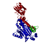
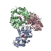
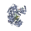
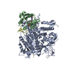
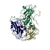
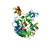
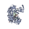
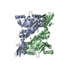
Crystal structure of C-terminal domain of Helicobacter pylori DnaB Helicase
Components
REPLICATIVE DNA HELICASE
Keywords
HYDROLASE / DNA REPLICATION / ATPASE
Function / homology
Function and homology information
primosome complex / DNA replication, synthesis of primer / DNA 5'-3' helicase / DNA helicase activity / 5'-3' DNA helicase activity / DNA replication / ATP hydrolysis activity / DNA binding / ATP binding / identical protein binding / cytosol Similarity search - Function
DNA helicase, DnaB type / DNA helicase, DnaB-like, N-terminal / DnaB-like helicase N terminal domain / DNA helicase, DnaB-like, N-terminal domain superfamily / DNA helicase DnaB, N-terminal/DNA primase DnaG, C-terminal / DnaB-like helicase C terminal domain / DNA helicase, DnaB-like, C-terminal / Superfamily 4 helicase domain profile. / P-loop containing nucleotide triphosphate hydrolases / Rossmann fold ...DNA helicase, DnaB type / DNA helicase, DnaB-like, N-terminal / DnaB-like helicase N terminal domain / DNA helicase, DnaB-like, N-terminal domain superfamily / DNA helicase DnaB, N-terminal/DNA primase DnaG, C-terminal / DnaB-like helicase C terminal domain / DNA helicase, DnaB-like, C-terminal / Superfamily 4 helicase domain profile. / P-loop containing nucleotide triphosphate hydrolases / Rossmann fold / P-loop containing nucleoside triphosphate hydrolase / 3-Layer(aba) Sandwich / Alpha Beta Similarity search - Domain/homology
 Movie
Movie Controller

 Yorodumi
 Open data
 Basic information
 Components
 Keywords
 Function and homology information
 HELICOBACTER PYLORI (bacteria)
 X-RAY DIFFRACTION /
  Authors
 Citation
 Structure visualization
 Downloads & links
 Other downloads
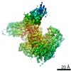

 PDBj
PDBj


 Assembly
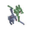

 Mass: 189.100 Da / Num. of mol.: 3 / Source method: obtained synthetically / Formula: C6H5O7
 Mass: 18.015 Da / Num. of mol.: 105 / Source method: isolated from a natural source / Formula: H2O
 Sample preparation
 / Beamline: ID14-4 / Wavelength: 0.9
  Processing