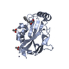
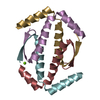
- PDB-3zy1: Crystal structure of the human p63 tetramerization domain -
+
Open data
ID or keywords:
Loading...
-
Basic information
Entry
Database: PDB / ID: 3zy1
Title
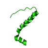
Crystal structure of the human p63 tetramerization domain
Components
TUMOR PROTEIN 63
Keywords
TRANSCRIPTION / TRANSCRIPTION FACTOR / TETRAMERIZATION DOMAIN / CELL-CYCLE CONTROL
Function / homology
Function and homology information
positive regulation of somatic stem cell population maintenance / positive regulation of fibroblast apoptotic process / negative regulation of intracellular estrogen receptor signaling pathway / positive regulation of cell cycle G1/S phase transition / Developmental Lineage of Mammary Stem Cells / Differentiation of Keratinocytes in Interfollicular Epidermis in Mammalian Skin / negative regulation of cellular senescence / TP53 Regulates Transcription of Death Receptors and Ligands / Activation of PUMA and translocation to mitochondria / Regulation of TP53 Activity through Association with Co-factors ...positive regulation of somatic stem cell population maintenance / positive regulation of fibroblast apoptotic process / negative regulation of intracellular estrogen receptor signaling pathway / positive regulation of cell cycle G1/S phase transition / Developmental Lineage of Mammary Stem Cells / Differentiation of Keratinocytes in Interfollicular Epidermis in Mammalian Skin / negative regulation of cellular senescence / TP53 Regulates Transcription of Death Receptors and Ligands / Activation of PUMA and translocation to mitochondria / Regulation of TP53 Activity through Association with Co-factors / WW domain binding / positive regulation of Notch signaling pathway / TP53 Regulates Transcription of Caspase Activators and Caspases / regulation of epidermal cell division / TP53 Regulates Transcription of Genes Involved in Cytochrome C Release / Developmental Lineage of Mammary Gland Myoepithelial Cells / positive regulation of osteoblast differentiation / TP53 regulates transcription of several additional cell death genes whose specific roles in p53-dependent apoptosis remain uncertain / intrinsic apoptotic signaling pathway in response to DNA damage by p53 class mediator / establishment of skin barrier / Developmental Lineage of Mammary Gland Luminal Epithelial Cells / Notch signaling pathway / Pyroptosis / MDM2/MDM4 family protein binding / TP53 Regulates Metabolic Genes / RNA polymerase II transcription regulatory region sequence-specific DNA binding / protein tetramerization / promoter-specific chromatin binding / p53 binding / spermatogenesis / regulation of apoptotic process / DNA-binding transcription factor activity, RNA polymerase II-specific / RNA polymerase II cis-regulatory region sequence-specific DNA binding / DNA-binding transcription factor activity / negative regulation of DNA-templated transcription / apoptotic process / positive regulation of cell population proliferation / chromatin binding / DNA damage response / dendrite / positive regulation of DNA-templated transcription / chromatin / positive regulation of transcription by RNA polymerase II / protein-containing complex / DNA binding / DNA-templated transcription / nucleoplasm / metal ion binding / identical protein binding / nucleus / cytoplasm Similarity search - Function
TUMORPROTEIN63 / P63 / CHRONIC ULCERATIVE STOMATITIS PROTEIN / CUSP / KERATINOCYTE TRANSCRIPTION FACTOR KET / ...P63 / CHRONIC ULCERATIVE STOMATITIS PROTEIN / CUSP / KERATINOCYTE TRANSCRIPTION FACTOR KET / TRANSFORMATION-RELATED PROTEIN 63 / TP63 / TUMOR PROTEIN P73-LIKE / P73L / P40 / P51
Mass: 5604.372 Da / Num. of mol.: 1 / Fragment: TETRAMERIZATION DOMAIN, RESIDUES 398-441 Source method: isolated from a genetically manipulated source Source: (gene. exp.) HOMO SAPIENS (human) / Production host: ESCHERICHIA COLI (E. coli) / References: UniProt: Q9H3D4
-
Experimental details
-
Experiment
Experiment
Method: X-RAY DIFFRACTION
-
Sample preparation
Crystal
Density Matthews: 3 Å3/Da / Density % sol: 59 % / Description: NONE
Crystal grow
Temperature: 290 K / Method: vapor diffusion, sitting drop Details: SITTING DROP VAPOR DIFFUSION AT 17 DEGREE C. PROTEIN SOLUTION: 12-15 MG/ML IN 20 MM TRIS PH 8.5, 50 MM NACL CRYSTALLIZATION BUFFER: 10% PEG 8000, 0.1 M HEPES PH 7.5, AND 0.2 M CA ACETATE
 Movie
Movie Controller


 Open data
 Basic information
 Components
 Keywords
 Function and homology information
 HOMO SAPIENS (human)
 X-RAY DIFFRACTION /
  Authors
 Citation
 Structure visualization
 Downloads & links
 Other downloads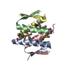


 PDBj
PDBj





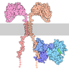
 Assembly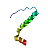

 ESCHERICHIA COLI (E. coli) / References: UniProt: Q9H3D4
 Sample preparation
 / Beamline: I03 / Wavelength: 0.9791
  Processing