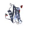
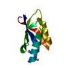
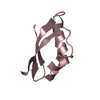
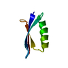
[English] 日本語
Yorodumi
- PDB-2y9u: Structural basis of p63a SAM domain mutants involved in AEC syndrome -

+
Open data


ID or keywords:

Loading...

-
Basic information

Entry
Database: PDB / ID: 2y9u
TitleStructural basis of p63a SAM domain mutants involved in AEC syndrome
ComponentsTUMOR PROTEIN 63
KeywordsAPOPTOSIS / STERILE ALPHA MOTIF / 5-HELIX BUNDLE / MUTATIONS / AEC SYNDROME
Function / homology
Function and homology information


ectoderm and mesoderm interaction / epidermal cell division / cloacal septation / positive regulation of somatic stem cell population maintenance / prostatic bud formation / negative regulation of mesoderm development / female genitalia morphogenesis / establishment of planar polarity / positive regulation of keratinocyte proliferation / negative regulation of keratinocyte differentiation ...ectoderm and mesoderm interaction / epidermal cell division / cloacal septation / positive regulation of somatic stem cell population maintenance / prostatic bud formation / negative regulation of mesoderm development / female genitalia morphogenesis / establishment of planar polarity / positive regulation of keratinocyte proliferation / negative regulation of keratinocyte differentiation / squamous basal epithelial stem cell differentiation involved in prostate gland acinus development / polarized epithelial cell differentiation / negative regulation of intracellular estrogen receptor signaling pathway / proximal/distal pattern formation / positive regulation of fibroblast apoptotic process / positive regulation of cell cycle G1/S phase transition / WW domain binding / skin morphogenesis / cranial skeletal system development / sympathetic nervous system development / post-anal tail morphogenesis / embryonic forelimb morphogenesis / embryonic hindlimb morphogenesis / TP53 Regulates Transcription of Death Receptors and Ligands / Activation of PUMA and translocation to mitochondria / Regulation of TP53 Activity through Association with Co-factors / hair follicle morphogenesis / positive regulation of Notch signaling pathway / regulation of epidermal cell division / TP53 regulates transcription of several additional cell death genes whose specific roles in p53-dependent apoptosis remain uncertain / positive regulation of stem cell proliferation / epithelial cell development / TP53 Regulates Transcription of Caspase Activators and Caspases / odontogenesis of dentin-containing tooth / negative regulation of cellular senescence / TP53 Regulates Transcription of Genes Involved in Cytochrome C Release / keratinocyte proliferation / intrinsic apoptotic signaling pathway in response to DNA damage by p53 class mediator / establishment of skin barrier / Pyroptosis / positive regulation of osteoblast differentiation / keratinocyte differentiation / Notch signaling pathway / MDM2/MDM4 family protein binding / stem cell proliferation / skeletal system development / determination of adult lifespan / promoter-specific chromatin binding / TP53 Regulates Metabolic Genes / positive regulation of apoptotic signaling pathway / protein tetramerization / RNA polymerase II transcription regulatory region sequence-specific DNA binding / cellular senescence / p53 binding / spermatogenesis / DNA-binding transcription activator activity, RNA polymerase II-specific / neuron apoptotic process / transcription by RNA polymerase II / damaged DNA binding / DNA-binding transcription factor activity, RNA polymerase II-specific / chromatin remodeling / RNA polymerase II cis-regulatory region sequence-specific DNA binding / DNA-binding transcription factor activity / negative regulation of DNA-templated transcription / apoptotic process / DNA damage response / chromatin binding / dendrite / chromatin / regulation of transcription by RNA polymerase II / positive regulation of DNA-templated transcription / negative regulation of transcription by RNA polymerase II / positive regulation of transcription by RNA polymerase II / DNA binding / nucleoplasm / identical protein binding / nucleus / metal ion binding / cytoplasm
Similarity search - Function
Tumour protein p63, SAM domain / Transcription Factor, Ets-1 / p53 family signature. / p53, tetramerisation domain / P53 tetramerisation motif / p53, DNA-binding domain / P53 DNA-binding domain / p53 tumour suppressor family / p53-like tetramerisation domain superfamily / p53/RUNT-type transcription factor, DNA-binding domain superfamily ...Tumour protein p63, SAM domain / Transcription Factor, Ets-1 / p53 family signature. / p53, tetramerisation domain / P53 tetramerisation motif / p53, DNA-binding domain / P53 DNA-binding domain / p53 tumour suppressor family / p53-like tetramerisation domain superfamily / p53/RUNT-type transcription factor, DNA-binding domain superfamily / SAM domain (Sterile alpha motif) / p53-like transcription factor, DNA-binding / Sterile alpha motif. / Sterile alpha motif domain / Sterile alpha motif/pointed domain superfamily / DNA polymerase; domain 1 / Orthogonal Bundle / Mainly Alpha
Similarity search - Domain/homology
Biological speciesHOMO SAPIENS (human)
MethodX-RAY DIFFRACTION / SYNCHROTRON / MOLECULAR REPLACEMENT / Resolution: 1.6 Å
AuthorsSathyamurthy, A. / Freund, S.M.V. / Johnson, C.M. / Allen, M.D.
CitationJournal: FEBS J. / Year: 2011
Title: Structural Basis of P63Alpha Sam Domain Mutants Involved in Aec Syndrome.
Authors: Sathyamurthy, A. / Freund, S.M.V. / Johnson, C.M. / Allen, M.D. / Bycroft, M.
History
DepositionFeb 16, 2011Deposition site: PDBE / Processing site: PDBE
Revision 1.0Aug 3, 2011Provider: repository / Type: Initial release
Revision 1.1Jan 24, 2018Group: Source and taxonomy / Category: entity_src_gen
Item: _entity_src_gen.pdbx_host_org_ncbi_taxonomy_id / _entity_src_gen.pdbx_host_org_scientific_name ..._entity_src_gen.pdbx_host_org_ncbi_taxonomy_id / _entity_src_gen.pdbx_host_org_scientific_name / _entity_src_gen.pdbx_host_org_strain / _entity_src_gen.pdbx_host_org_variant
Revision 1.2May 8, 2024Group: Data collection / Database references ...Data collection / Database references / Derived calculations / Other
Category: chem_comp_atom / chem_comp_bond ...chem_comp_atom / chem_comp_bond / database_2 / pdbx_database_status / struct_site
Item: _database_2.pdbx_DOI / _database_2.pdbx_database_accession ..._database_2.pdbx_DOI / _database_2.pdbx_database_accession / _pdbx_database_status.status_code_sf / _struct_site.pdbx_auth_asym_id / _struct_site.pdbx_auth_comp_id / _struct_site.pdbx_auth_seq_id

-
Structure visualization

Structure viewerMolecule:
MolmilJmol/JSmol

Downloads & links

-
Assembly

Deposited unit
A: TUMOR PROTEIN 63
hetero molecules


Theoretical massNumber of molelcules
Total (without water)8,0012
Polymers7,9051
Non-polymers961
Water1,45981
1


  • Idetical with deposited unit
  • defined by author&software
TypeNameSymmetry operationNumber
identity operation1_555x,y,z1
MethodPISA
Unit cell
Length a, b, c (Å)34.624, 38.309, 44.516
Angle α, β, γ (deg.)90.00, 90.00, 90.00
Int Tables number19
Space group name H-MP212121

-
Components

#1: Protein TUMOR PROTEIN 63 / CHRONIC ULCERATIVE STOMATITIS PROTEIN / KERATINOCYTE TRANSCRIPTION FACTOR KET / TRANSFORMATION- ...CHRONIC ULCERATIVE STOMATITIS PROTEIN / KERATINOCYTE TRANSCRIPTION FACTOR KET / TRANSFORMATION-RELATED PROTEIN 63 / TUMOR PROTEIN P73-LIKE / P40 / P51 / P63 / CUSP / TP63 / P73L


Mass: 7904.923 Da / Num. of mol.: 1 / Fragment: SAM DOMAIN, RESIDUES 545-611
Source method: isolated from a genetically manipulated source
Source: (gene. exp.) HOMO SAPIENS (human) / Production host: ESCHERICHIA COLI BL21(DE3) (bacteria) / Variant (production host): C41 / References: UniProt: Q9H3D4
#2: Chemical ChemComp-SO4 / SULFATE ION


Mass: 96.063 Da / Num. of mol.: 1 / Source method: obtained synthetically / Formula: SO4
#3: Water ChemComp-HOH / water


Mass: 18.015 Da / Num. of mol.: 81 / Source method: isolated from a natural source / Formula: H2O
Sequence detailsGLY AND SER AND THE N-TERMINUS ARE FROM THE FUSION TAG

-
Experimental details

-
Experiment

ExperimentMethod: X-RAY DIFFRACTION / Number of used crystals: 1

-
Sample preparation

CrystalDensity Matthews: 1.87 Å3/Da / Density % sol: 34.13 % / Description: NONE
Crystal growpH: 6.4
Details: 100 MM SODIUM CITRATE, PH 6.4, 500MM LITHIUM SULPHATE, 500MM AMMONIUM SULPHATE, 5MM DTT

-
Data collection

DiffractionMean temperature: 100 K
Diffraction sourceSource: SYNCHROTRON / Site: ESRF / Beamline: ID14-2 / Wavelength: 0.9795
DetectorType: ADSC CCD / Detector: CCD
RadiationProtocol: SINGLE WAVELENGTH / Monochromatic (M) / Laue (L): M / Scattering type: x-ray
Radiation wavelengthWavelength: 0.9795 Å / Relative weight: 1
ReflectionResolution: 1.6→27.4 Å / Num. obs: 8205 / % possible obs: 98.9 % / Observed criterion σ(I): 2 / Redundancy: 5.3 % / Biso Wilson estimate: 15.69 Å2 / Rmerge(I) obs: 0.11 / Net I/σ(I): 14
Reflection shellHighest resolution: 1.6 Å / Redundancy: 5.5 % / Rmerge(I) obs: 0.3 / Mean I/σ(I) obs: 4.7 / % possible all: 96

-
Processing

Software
NameVersionClassification
PHENIX(PHENIX.REFINE)refinement
MOSFLMdata reduction
SCALAdata scaling
PHASERphasing
RefinementMethod to determine structure: MOLECULAR REPLACEMENT / Resolution: 1.6→22.249 Å / SU ML: 0.16 / σ(F): 1.32 / Phase error: 19.05 / Stereochemistry target values: ML
RfactorNum. reflection% reflection
Rfree0.2056 757 5.2 %
Rwork0.1853 --
obs0.1863 8175 97.23 %
Solvent computationShrinkage radii: 0.9 Å / VDW probe radii: 1.11 Å / Solvent model: FLAT BULK SOLVENT MODEL / Bsol: 50.799 Å2 / ksol: 0.399 e/Å3
Displacement parameters
Baniso -1Baniso -2Baniso -3
1-4.5086 Å20 Å20 Å2
2--1.1207 Å20 Å2
3----6.7947 Å2
Refinement stepCycle: LAST / Resolution: 1.6→22.249 Å
ProteinNucleic acidLigandSolventTotal
Num. atoms544 0 5 81 630
Refine LS restraints
Refine-IDTypeDev idealNumber
X-RAY DIFFRACTIONf_bond_d0.009562
X-RAY DIFFRACTIONf_angle_d1.192761
X-RAY DIFFRACTIONf_dihedral_angle_d16.183194
X-RAY DIFFRACTIONf_chiral_restr0.07384
X-RAY DIFFRACTIONf_plane_restr0.00595
LS refinement shell
Resolution (Å)Rfactor RfreeNum. reflection RfreeRfactor RworkNum. reflection RworkRefine-ID% reflection obs (%)
1.6001-1.72360.2251660.20022782X-RAY DIFFRACTION97
1.7236-1.89690.20171670.18492777X-RAY DIFFRACTION97
1.8969-2.17120.22141500.17682794X-RAY DIFFRACTION97
2.1712-2.73470.23061360.18122793X-RAY DIFFRACTION97
2.7347-22.25090.18171380.18382782X-RAY DIFFRACTION97

+
About Yorodumi

-
News

-
Feb 9, 2022. New format data for meta-information of EMDB entries

New format data for meta-information of EMDB entries

  • Version 3 of the EMDB header file is now the official format.
  • The previous official version 1.9 will be removed from the archive.

Related info.:EMDB header

External links:wwPDB to switch to version 3 of the EMDB data model

-
Aug 12, 2020. Covid-19 info

Covid-19 info

URL: https://pdbjlvh1.pdbj.org/emnavi/covid19.php

New page: Covid-19 featured information page in EM Navigator.

Related info.:Covid-19 info / Mar 5, 2020. Novel coronavirus structure data

+
Mar 5, 2020. Novel coronavirus structure data

Novel coronavirus structure data

Related info.:Yorodumi Speices / Aug 12, 2020. Covid-19 info

External links:COVID-19 featured content - PDBj / Molecule of the Month (242):Coronavirus Proteases

+
Jan 31, 2019. EMDB accession codes are about to change! (news from PDBe EMDB page)

EMDB accession codes are about to change! (news from PDBe EMDB page)

  • The allocation of 4 digits for EMDB accession codes will soon come to an end. Whilst these codes will remain in use, new EMDB accession codes will include an additional digit and will expand incrementally as the available range of codes is exhausted. The current 4-digit format prefixed with “EMD-” (i.e. EMD-XXXX) will advance to a 5-digit format (i.e. EMD-XXXXX), and so on. It is currently estimated that the 4-digit codes will be depleted around Spring 2019, at which point the 5-digit format will come into force.
  • The EM Navigator/Yorodumi systems omit the EMD- prefix.

Related info.:Q: What is EMD? / ID/Accession-code notation in Yorodumi/EM Navigator

External links:EMDB Accession Codes are Changing Soon! / Contact to PDBj

+
Jul 12, 2017. Major update of PDB

Major update of PDB

  • wwPDB released updated PDB data conforming to the new PDBx/mmCIF dictionary.
  • This is a major update changing the version number from 4 to 5, and with Remediation, in which all the entries are updated.
  • In this update, many items about electron microscopy experimental information are reorganized (e.g. em_software).
  • Now, EM Navigator and Yorodumi are based on the updated data.

External links:wwPDB Remediation / Enriched Model Files Conforming to OneDep Data Standards Now Available in the PDB FTP Archive

-
Yorodumi

Thousand views of thousand structures

  • Yorodumi is a browser for structure data from EMDB, PDB, SASBDB, etc.
  • This page is also the successor to EM Navigator detail page, and also detail information page/front-end page for Omokage search.
  • The word "yorodu" (or yorozu) is an old Japanese word meaning "ten thousand". "mi" (miru) is to see.

Related info.:EMDB / PDB / SASBDB / Comparison of 3 databanks / Yorodumi Search / Aug 31, 2016. New EM Navigator & Yorodumi / Yorodumi Papers / Jmol/JSmol / Function and homology information / Changes in new EM Navigator and Yorodumi

Read more