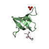
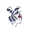
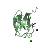
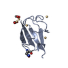
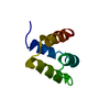
- PDB-1rg6: Solution structure of the C-terminal domain of p63 -
+
Open data
ID or keywords:
Loading...
-
Basic information
Entry
Database: PDB / ID: 1rg6
Title
Solution structure of the C-terminal domain of p63
Components
second splice variant p63
Keywords
GENE REGULATION / P73 SAM-LIKE DOMAIN
Function / homology
Function and homology information
positive regulation of somatic stem cell population maintenance / positive regulation of fibroblast apoptotic process / negative regulation of intracellular estrogen receptor signaling pathway / Developmental Lineage of Mammary Gland Luminal Epithelial Cells / positive regulation of cell cycle G1/S phase transition / Developmental Lineage of Mammary Stem Cells / Differentiation of Keratinocytes in Interfollicular Epidermis in Mammalian Skin / TP53 Regulates Transcription of Death Receptors and Ligands / Activation of PUMA and translocation to mitochondria / Regulation of TP53 Activity through Association with Co-factors ...positive regulation of somatic stem cell population maintenance / positive regulation of fibroblast apoptotic process / negative regulation of intracellular estrogen receptor signaling pathway / Developmental Lineage of Mammary Gland Luminal Epithelial Cells / positive regulation of cell cycle G1/S phase transition / Developmental Lineage of Mammary Stem Cells / Differentiation of Keratinocytes in Interfollicular Epidermis in Mammalian Skin / TP53 Regulates Transcription of Death Receptors and Ligands / Activation of PUMA and translocation to mitochondria / Regulation of TP53 Activity through Association with Co-factors / WW domain binding / positive regulation of Notch signaling pathway / TP53 Regulates Transcription of Caspase Activators and Caspases / regulation of epidermal cell division / TP53 Regulates Transcription of Genes Involved in Cytochrome C Release / negative regulation of cellular senescence / Developmental Lineage of Mammary Gland Myoepithelial Cells / TP53 regulates transcription of several additional cell death genes whose specific roles in p53-dependent apoptosis remain uncertain / establishment of skin barrier / positive regulation of osteoblast differentiation / intrinsic apoptotic signaling pathway in response to DNA damage by p53 class mediator / Pyroptosis / Notch signaling pathway / MDM2/MDM4 family protein binding / TP53 Regulates Metabolic Genes / RNA polymerase II transcription regulatory region sequence-specific DNA binding / protein tetramerization / promoter-specific chromatin binding / p53 binding / regulation of apoptotic process / spermatogenesis / DNA-binding transcription factor activity, RNA polymerase II-specific / RNA polymerase II cis-regulatory region sequence-specific DNA binding / DNA-binding transcription factor activity / negative regulation of DNA-templated transcription / chromatin binding / apoptotic process / positive regulation of cell population proliferation / DNA damage response / dendrite / positive regulation of DNA-templated transcription / chromatin / DNA-templated transcription / positive regulation of transcription by RNA polymerase II / protein-containing complex / DNA binding / nucleoplasm / metal ion binding / identical protein binding / nucleus / cytoplasm Similarity search - Function
In the structure databanks used in Yorodumi, some data are registered as the other names, "COVID-19 virus" and "2019-nCoV". Here are the details of the virus and the list of structure data.
Jan 31, 2019. EMDB accession codes are about to change! (news from PDBe EMDB page)
EMDB accession codes are about to change! (news from PDBe EMDB page)
The allocation of 4 digits for EMDB accession codes will soon come to an end. Whilst these codes will remain in use, new EMDB accession codes will include an additional digit and will expand incrementally as the available range of codes is exhausted. The current 4-digit format prefixed with “EMD-” (i.e. EMD-XXXX) will advance to a 5-digit format (i.e. EMD-XXXXX), and so on. It is currently estimated that the 4-digit codes will be depleted around Spring 2019, at which point the 5-digit format will come into force.
The EM Navigator/Yorodumi systems omit the EMD- prefix.
Related info.:Q: What is EMD? / ID/Accession-code notation in Yorodumi/EM Navigator
Yorodumi is a browser for structure data from EMDB, PDB, SASBDB, etc.
This page is also the successor to EM Navigator detail page, and also detail information page/front-end page for Omokage search.
The word "yorodu" (or yorozu) is an old Japanese word meaning "ten thousand". "mi" (miru) is to see.
Related info.:EMDB / PDB / SASBDB / Comparison of 3 databanks / Yorodumi Search / Aug 31, 2016. New EM Navigator & Yorodumi / Yorodumi Papers / Jmol/JSmol / Function and homology information / Changes in new EM Navigator and Yorodumi
 Movie
Movie Controller


 Open data
 Basic information
 Components
 Keywords
 Function and homology information
 Homo sapiens (human)
 Authors
 Citation
 Structure visualization
 Downloads & links
 Other downloads
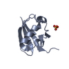

 PDBj
PDBj


 Assembly

 HSQC
HSQC Sample preparation
 Processing