 Movie
Movie Controller

[English] 日本語
 Yorodumi
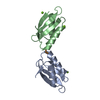
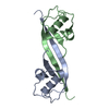
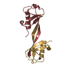
- PDB-2qif: Crystal structure of a metallochaperone with a tetranuclear Cu(I)... -
+ Open data

- Basic information
| Entry | Database: PDB / ID: 2qif | ||||||
|---|---|---|---|---|---|---|---|
| Title | Crystal structure of a metallochaperone with a tetranuclear Cu(I) cluster | ||||||
 Components Components | Copper chaperone copZ | ||||||
 Keywords Keywords | CHAPERONE / TETRANUCLEAR CU(I) CLUSTER | ||||||
| Function / homology |  Function and homology information Function and homology information | ||||||
| Biological species |  | ||||||
| Method |  X-RAY DIFFRACTION / SYNCHROTRON / SAD / Resolution: 1.5 Å X-RAY DIFFRACTION / SYNCHROTRON / SAD / Resolution: 1.5 Å | ||||||
 Authors Authors | West, C. / Singleton, C. / Kihlken, M.A. / Le Brun, N.E. / Hemmings, A.M. | ||||||
 Citation Citation | Journal: Biochemistry / Year: 2009 Title: A tetranuclear Cu(I) cluster in the metallochaperone protein CopZ. Authors: Hearnshaw, S. / West, C. / Singleton, C. / Zhou, L. / Kihlken, M.A. / Strange, R.W. / Le Brun, N.E. / Hemmings, A.M. #1: Journal: Biochem.J. / Year: 2002 Title: Copper-mediated dimerization of CopZ, a predicted copper chaperone from Bacillus subtilis Authors: Kihlken, M.A. / Leech, A.P. / Le Brun, N.E. | ||||||
| History |
|
- Structure visualization
| Structure viewer | Molecule: MolmilJmol/JSmol |
|---|
- Downloads & links
-Download
| PDBx/mmCIF format | 2qif.cif.gz | 75.9 KB | Display | PDBx/mmCIF format |
|---|---|---|---|---|
| PDB format | pdb2qif.ent.gz | 56.3 KB | Display | PDB format |
| PDBx/mmJSON format | 2qif.json.gz | Tree view | PDBx/mmJSON format | |
| Others |  Other downloads Other downloads |
-Validation report
| Arichive directory | https://data.pdbj.org/pub/pdb/validation_reports/qi/2qifftp://data.pdbj.org/pub/pdb/validation_reports/qi/2qif | HTTPS FTP |
|---|
-Related structure data
| Related structure data | |
|---|---|
| Similar structure data |


-Links
 PDBj
PDBj
- Assembly
| Deposited unit | 
| ||||||||
|---|---|---|---|---|---|---|---|---|---|
| 1 |
| ||||||||
| 2 | 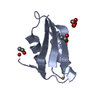
| ||||||||
| 3 | 
| ||||||||
| Unit cell |
| ||||||||
| Details | THE CRYSTALLOGRAPHIC ASYMMETRIC UNIT CONTAINS A SINGLE COPY OF THE BIOLOGICAL UNIT |
-Components
-Protein , 1 types, 2 molecules AB
| #1: Protein | Mass: 7346.172 Da / Num. of mol.: 2 Source method: isolated from a genetically manipulated source Source: (gene. exp.) |
|---|
-Non-polymers , 5 types, 174 molecules 


| #2: Chemical | ChemComp-CU1 /  Mass: 63.546 Da / Num. of mol.: 4 / Source method: obtained synthetically / Formula: Cu Mass: 63.546 Da / Num. of mol.: 4 / Source method: obtained synthetically / Formula: Cu#3: Chemical |  Mass: 59.044 Da / Num. of mol.: 2 / Source method: obtained synthetically / Formula: C2H3O2 Mass: 59.044 Da / Num. of mol.: 2 / Source method: obtained synthetically / Formula: C2H3O2#4: Chemical | ChemComp-GOL / |  Mass: 92.094 Da / Num. of mol.: 1 / Source method: obtained synthetically / Formula: C3H8O3 Mass: 92.094 Da / Num. of mol.: 1 / Source method: obtained synthetically / Formula: C3H8O3#5: Chemical | ChemComp-CA / |  Mass: 40.078 Da / Num. of mol.: 1 / Source method: obtained synthetically / Formula: Ca Mass: 40.078 Da / Num. of mol.: 1 / Source method: obtained synthetically / Formula: Ca#6: Water | ChemComp-HOH / | Mass: 18.015 Da / Num. of mol.: 166 / Source method: isolated from a natural source / Formula: H2O |
|---|
-Experimental details
-Experiment
| Experiment | Method: X-RAY DIFFRACTION / Number of used crystals: 1 |
|---|
- Sample preparation
| Crystal | Density Matthews: 1.98 Å3/Da / Density % sol: 49.52 % |
|---|---|
| Crystal grow | Temperature: 277 K / Method: vapor batch / pH: 4.6 Details: Crystallization by vapour batch using 12 microL drops in Terasaki plates covered with a thin layer of silicone oil and equilibrated against 10 % (v/v) isopropanol (24 h) then 20% (v/v) ...Details: Crystallization by vapour batch using 12 microL drops in Terasaki plates covered with a thin layer of silicone oil and equilibrated against 10 % (v/v) isopropanol (24 h) then 20% (v/v) isopropanol for 4 days. 15% (v/v) glycerol added as cryoprotectant., pH 4.6, VAPOUR BATCH, temperature 277K |
-Data collection
| Diffraction | Mean temperature: 100 K |
|---|---|
| Diffraction source | Source: SYNCHROTRON / Site: SRS  / Beamline: PX14.2 / Wavelength: 0.98 Å / Beamline: PX14.2 / Wavelength: 0.98 Å |
| Detector | Type: ADSC QUANTUM 4 / Detector: CCD / Date: Oct 24, 2002 Details: mirror; vertical focusing, glancing angle 3.5 mrad, 7.0 Ang. cut off, 1.2m long silicon substrate, rhodium coated, distance from source 16m |
| Radiation | Monochromator: Si 111 optimised for 0.979 or 1.2 wavelength, horizontally focusing, distance from source 18m Protocol: SINGLE WAVELENGTH / Monochromatic (M) / Laue (L): M / Scattering type: x-ray |
| Radiation wavelength | Wavelength: 0.98 Å / Relative weight: 1 |
| Reflection | Resolution: 1.5→30 Å / Num. obs: 41550 / % possible obs: 94.3 % / Observed criterion σ(F): 0 / Observed criterion σ(I): 0 / Redundancy: 3.5 % / Biso Wilson estimate: 10.5 Å2 / Rmerge(I) obs: 0.038 / Rsym value: 0.038 / Χ2: 2.19 / Net I/σ(I): 25.7 |
| Reflection shell | Resolution: 1.5→1.55 Å / Redundancy: 2.9 % / Rmerge(I) obs: 0.147 / Mean I/σ(I) obs: 12.4 / Num. unique all: 3783 / Rsym value: 0.45 / Χ2: 2.636 / % possible all: 84.9 |
- Processing
| Software |
| |||||||||||||||||||||||||||||||||
|---|---|---|---|---|---|---|---|---|---|---|---|---|---|---|---|---|---|---|---|---|---|---|---|---|---|---|---|---|---|---|---|---|---|---|
| Refinement | Method to determine structure: SAD / Resolution: 1.5→10 Å / Num. parameters: 11523 / Num. restraintsaints: 14831 Isotropic thermal model: INDIVIDUAL ATOMIC ANISOTROPIC TEMPERATURE FACTOR REFINEMENT Cross valid method: FREE R / σ(F): 0 / σ(I): 0 / Stereochemistry target values: ENGH AND HUBER
| |||||||||||||||||||||||||||||||||
| Refine analyze | Num. disordered residues: 14 / Occupancy sum hydrogen: 0 / Occupancy sum non hydrogen: 1191.22 | |||||||||||||||||||||||||||||||||
| Refinement step | Cycle: LAST / Resolution: 1.5→10 Å
| |||||||||||||||||||||||||||||||||
| Refine LS restraints |
|
