 Movie
Movie Controller

[English] 日本語
 Yorodumi
- PDB-3zl4: Antibody structural organization: Role of kappa - lambda chain co... -
+ Open data

- Basic information
| Entry | Database: PDB / ID: 3zl4 | ||||||
|---|---|---|---|---|---|---|---|
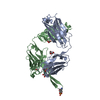
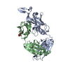
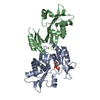
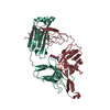
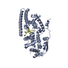
| Title | Antibody structural organization: Role of kappa - lambda chain constant domain switch in catalytic functionality | ||||||
 Components Components |
| ||||||
 Keywords Keywords | IMMUNE SYSTEM | ||||||
| Function / homology |  Function and homology information Function and homology informationimmunoglobulin complex / adaptive immune response / extracellular region / plasma membrane Similarity search - Function | ||||||
| Biological species |  Homo sapiens (human) Homo sapiens (human) | ||||||
| Method |  X-RAY DIFFRACTION / SYNCHROTRON / MOLECULAR REPLACEMENT / Resolution: 1.95 Å X-RAY DIFFRACTION / SYNCHROTRON / MOLECULAR REPLACEMENT / Resolution: 1.95 Å | ||||||
 Authors Authors | Chatziefthimiou, S.D. / Ponomarenko, N.A. / Kurkova, I.N. / Smirnov, A.V. / Smirnov, I.V. / Lamzin, V.S. / Gabibov, A.G. / Wilmanns, M. | ||||||
 Citation Citation | Journal: Acta Crystallogr.,Sect.D / Year: 2014 Title: Role of Kappa>Lambda Light-Chain Constant-Domain Switch in the Structure and Functionality of A17 Reactibody Authors: Ponomarenko, N.A. / Chatziefthimiou, S.D. / Kurkova, I.N. / Mokrushina, Y.A. / Stepanova, A.V. / Smirnov, I.V. / Avakyan, E.M. / Bobik, T.V. / Mamedov, A. / Mitkevich, V.A. / Belogurov, A.J. ...Authors: Ponomarenko, N.A. / Chatziefthimiou, S.D. / Kurkova, I.N. / Mokrushina, Y.A. / Stepanova, A.V. / Smirnov, I.V. / Avakyan, E.M. / Bobik, T.V. / Mamedov, A. / Mitkevich, V.A. / Belogurov, A.J. / Fedorova, O.S. / Dubina, M. / Golovin, A. / Lamzin, V.S. / Friboulet, A. / Makarov, A.A. / Wilmanns, M. / Gabibov, A.G. | ||||||
| History |
|
- Structure visualization
| Structure viewer | Molecule: MolmilJmol/JSmol |
|---|
- Downloads & links
-Download
| PDBx/mmCIF format | 3zl4.cif.gz | 104.2 KB | Display | PDBx/mmCIF format |
|---|---|---|---|---|
| PDB format | pdb3zl4.ent.gz | 79.3 KB | Display | PDB format |
| PDBx/mmJSON format | 3zl4.json.gz | Tree view | PDBx/mmJSON format | |
| Others |  Other downloads Other downloads |
-Validation report
| Arichive directory | https://data.pdbj.org/pub/pdb/validation_reports/zl/3zl4ftp://data.pdbj.org/pub/pdb/validation_reports/zl/3zl4 | HTTPS FTP |
|---|
-Related structure data
| Related structure data | 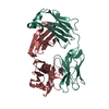 2xzaS S: Starting model for refinement |
|---|---|
| Similar structure data |


-Links
 PDBj
PDBj

- Assembly
| Deposited unit | 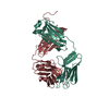
| ||||||||
|---|---|---|---|---|---|---|---|---|---|
| 1 |
| ||||||||
| Unit cell |
|
-Components
| #1: Antibody | Mass: 27415.270 Da / Num. of mol.: 1 Source method: isolated from a genetically manipulated source Source: (gene. exp.) Homo sapiens (human) / Plasmid: PPICZALFA / Production host:  KOMAGATAELLA PASTORIS GS115 (fungus) / References: UniProt: Q6GMX6*PLUS KOMAGATAELLA PASTORIS GS115 (fungus) / References: UniProt: Q6GMX6*PLUS |
|---|---|
| #2: Antibody | Mass: 26115.768 Da / Num. of mol.: 1 Source method: isolated from a genetically manipulated source Source: (gene. exp.) Homo sapiens (human) / Plasmid: PPICZALFA / Production host: KOMAGATAELLA PASTORIS GS115 (fungus) / References: UniProt: A2NUT2*PLUS |
| #3: Chemical | ChemComp-MES /   Mass: 195.237 Da / Num. of mol.: 1 / Source method: obtained synthetically / Formula: C6H13NO4S / Comment: pH buffer*YM Mass: 195.237 Da / Num. of mol.: 1 / Source method: obtained synthetically / Formula: C6H13NO4S / Comment: pH buffer*YM |
| #4: Water | ChemComp-HOH /  Mass: 18.015 Da / Num. of mol.: 310 / Source method: isolated from a natural source / Formula: H2O Mass: 18.015 Da / Num. of mol.: 310 / Source method: isolated from a natural source / Formula: H2O |
| Has protein modification | Y |
-Experimental details
-Experiment
| Experiment | Method: X-RAY DIFFRACTION / Number of used crystals: 1 |
|---|
- Sample preparation
| Crystal | Density Matthews: 2.8 Å3/Da / Density % sol: 55 % / Description: NONE |
|---|---|
| Crystal grow | pH: 6.5 Details: 0.1M MES PH 6.5, 0.25 M AMMONIUM SULFATE, 20% W/V PEG 5000 MME |
-Data collection
| Diffraction | Mean temperature: 100 K |
|---|---|
| Diffraction source | Source: SYNCHROTRON / Site: PETRA III, EMBL c/o DESY  / Beamline: P14 (MX2) / Wavelength: 1.2234 / Beamline: P14 (MX2) / Wavelength: 1.2234 |
| Detector | Type: MARRESEARCH / Detector: CCD / Date: May 14, 2012 |
| Radiation | Monochromator: SI(III) CRYSTAL MONOCHROMATOR / Protocol: SINGLE WAVELENGTH / Monochromatic (M) / Laue (L): M / Scattering type: x-ray |
| Radiation wavelength | Wavelength: 1.2234 Å / Relative weight: 1 |
| Reflection | Resolution: 1.95→25 Å / Num. obs: 39334 / % possible obs: 99.9 % / Observed criterion σ(I): 2 / Redundancy: 7.3 % / Rmerge(I) obs: 0.1 / Net I/σ(I): 15.6 |
| Reflection shell | Resolution: 1.95→2.06 Å / Redundancy: 7.3 % / Rmerge(I) obs: 0.85 / Mean I/σ(I) obs: 2.7 / % possible all: 100 |
- Processing
| Software |
| |||||||||||||||||||||||||||||||||||||||||||||||||||||||||||||||||||||||||||||||||||||||||||||||||||||||||
|---|---|---|---|---|---|---|---|---|---|---|---|---|---|---|---|---|---|---|---|---|---|---|---|---|---|---|---|---|---|---|---|---|---|---|---|---|---|---|---|---|---|---|---|---|---|---|---|---|---|---|---|---|---|---|---|---|---|---|---|---|---|---|---|---|---|---|---|---|---|---|---|---|---|---|---|---|---|---|---|---|---|---|---|---|---|---|---|---|---|---|---|---|---|---|---|---|---|---|---|---|---|---|---|---|---|---|
| Refinement | Method to determine structure: MOLECULAR REPLACEMENT Starting model: PDB ENTRY 2XZA Resolution: 1.95→24.399 Å / SU ML: 0.23 / σ(F): 1.38 / Phase error: 26.92 / Stereochemistry target values: ML
| |||||||||||||||||||||||||||||||||||||||||||||||||||||||||||||||||||||||||||||||||||||||||||||||||||||||||
| Solvent computation | Shrinkage radii: 0.9 Å / VDW probe radii: 1.11 Å / Solvent model: FLAT BULK SOLVENT MODEL | |||||||||||||||||||||||||||||||||||||||||||||||||||||||||||||||||||||||||||||||||||||||||||||||||||||||||
| Displacement parameters | Biso mean: 25.2 Å2 | |||||||||||||||||||||||||||||||||||||||||||||||||||||||||||||||||||||||||||||||||||||||||||||||||||||||||
| Refinement step | Cycle: LAST / Resolution: 1.95→24.399 Å
| |||||||||||||||||||||||||||||||||||||||||||||||||||||||||||||||||||||||||||||||||||||||||||||||||||||||||
| Refine LS restraints |
| |||||||||||||||||||||||||||||||||||||||||||||||||||||||||||||||||||||||||||||||||||||||||||||||||||||||||
| LS refinement shell |
|
