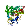
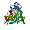
erythromycin 12-hydroxylase / macrolide biosynthetic process / oxidoreductase activity, acting on paired donors, with incorporation or reduction of molecular oxygen, NAD(P)H as one donor, and incorporation of one atom of oxygen / monooxygenase activity / NADP binding / iron ion binding / heme binding Similarity search - Function
Mass: 18.015 Da / Num. of mol.: 329 / Source method: isolated from a natural source / Formula: H2O
Sequence details
THE GENE USED FOR OBTAINING THE PROTEIN THAT WAS CRYSTALLIZED IS 15 AA LONGER AT THE NTERM WITH ...THE GENE USED FOR OBTAINING THE PROTEIN THAT WAS CRYSTALLIZED IS 15 AA LONGER AT THE NTERM WITH RESPECT TO THE UNIPROT GENE SEQUENCE P48635. ENGINEERED RESIDUE PHE 344 TO LEU.
-
Experimental details
-
Experiment
Experiment
Method: X-RAY DIFFRACTION / Number of used crystals: 1
-
Sample preparation
Crystal
Density Matthews: 1.91 Å3/Da / Density % sol: 34.97 %
Crystal grow
pH: 8.5 Details: 25% PEG3350, 0.1 M TRIS HCL, PH 8.5, 0.2 M CH3COONH4
Resolution: 2→29.12 Å / Cor.coef. Fo:Fc: 0.954 / Cor.coef. Fo:Fc free: 0.928 / SU B: 3.872 / SU ML: 0.11 / Cross valid method: THROUGHOUT / ESU R: 0.2 / ESU R Free: 0.169 / Stereochemistry target values: MAXIMUM LIKELIHOOD / Details: HYDROGENS HAVE BEEN ADDED IN THE RIDING POSITIONS.
Rfactor
Num. reflection
% reflection
Selection details
Rfree
0.21529
1367
5 %
RANDOM
Rwork
0.16332
-
-
-
obs
0.16597
25901
98.15 %
-
Solvent computation
Ion probe radii: 0.8 Å / Shrinkage radii: 0.8 Å / VDW probe radii: 1.2 Å / Solvent model: MASK
 Movie
Movie Controller

 Yorodumi
 Open data
 Basic information
 Components
 Keywords
 Function and homology information
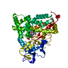
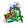
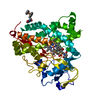
 SACCHAROPOLYSPORA ERYTHRAEA NRRL 2338 (bacteria)
 X-RAY DIFFRACTION /
  Authors
 Citation
 Structure visualization
 Downloads & links
 Other downloads
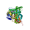


 PDBj
PDBj
 Assembly

 Mass: 616.487 Da / Num. of mol.: 1 / Source method: obtained synthetically / Formula: C34H32FeN4O4
 Mass: 717.927 Da / Num. of mol.: 1 / Source method: obtained synthetically / Formula: C37H67NO12
 Mass: 18.015 Da / Num. of mol.: 329 / Source method: isolated from a natural source / Formula: H2O
 Sample preparation
 / Beamline: ID23-2 / Wavelength: 0.873
  Processing