 Movie
Movie Controller


+ Open data

- Basic information
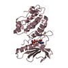
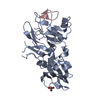
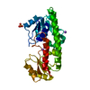
| Entry | Database: PDB / ID: 3zde | ||||||
|---|---|---|---|---|---|---|---|
| Title | Potassium free structure of E. coli ExoIX | ||||||
 Components Components | PROTEIN XNI | ||||||
 Keywords Keywords | HYDROLASE / ENDONUCLEASE / DNA BINDING | ||||||
| Function / homology |  Function and homology information Function and homology informationDNA replication, Okazaki fragment processing / 5'-flap endonuclease activity / 5'-3' exonuclease activity / potassium ion binding / Hydrolases; Acting on ester bonds / magnesium ion binding / DNA binding Similarity search - Function | ||||||
| Biological species |  | ||||||
| Method |  X-RAY DIFFRACTION / MOLECULAR REPLACEMENT / Resolution: 2.45 Å X-RAY DIFFRACTION / MOLECULAR REPLACEMENT / Resolution: 2.45 Å | ||||||
 Authors Authors | Hemsworth, G.R. / Anstey-Gilbert, C.S. / Flemming, C.S. / Hodskinson, M.R.G. / Zhang, J. / Sedelnikova, S.E. / Stillman, T.J. / Sayers, J.R. / Artymiuk, P.J. | ||||||
 Citation Citation | Journal: Nucleic Acids Res. / Year: 2013 Title: The Structure of E. Coli Exoix - Implications for DNA Binding and Catalysis in Flap Endonucleases Authors: Anstey-Gilbert, C.S. / Hemsworth, G.R. / Flemming, C.S. / Hodskinson, M.R.G. / Zhang, J. / Sedelnikova, S.E. / Stillman, T.J. / Sayers, J.R. / Artymiuk, P.J. | ||||||
| History |
|
- Structure visualization
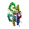
| Structure viewer | Molecule: MolmilJmol/JSmol |
|---|
- Downloads & links
-Download
| PDBx/mmCIF format | 3zde.cif.gz | 59.2 KB | Display | PDBx/mmCIF format |
|---|---|---|---|---|
| PDB format | pdb3zde.ent.gz | 43.4 KB | Display | PDB format |
| PDBx/mmJSON format | 3zde.json.gz | Tree view | PDBx/mmJSON format | |
| Others |  Other downloads Other downloads |
-Validation report
| Arichive directory | https://data.pdbj.org/pub/pdb/validation_reports/zd/3zdeftp://data.pdbj.org/pub/pdb/validation_reports/zd/3zde | HTTPS FTP |
|---|
-Related structure data
| Related structure data |  3zd8C  3zd9C  3zdaC  3zdbC  3zdcC  3zddC C: citing same article ( |
|---|---|
| Similar structure data |


-Links
 PDBj
PDBj

- Assembly
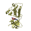
| Deposited unit | 
| ||||||||
|---|---|---|---|---|---|---|---|---|---|
| 1 |
| ||||||||
| Unit cell |
|
-Components
| #1: Protein | Mass: 28203.158 Da / Num. of mol.: 1 Source method: isolated from a genetically manipulated source Source: (gene. exp.) References: UniProt: Q8X6R9, Hydrolases; Acting on ester bonds |
|---|---|
| #2: Water | ChemComp-HOH /  Mass: 18.015 Da / Num. of mol.: 17 / Source method: isolated from a natural source / Formula: H2O Mass: 18.015 Da / Num. of mol.: 17 / Source method: isolated from a natural source / Formula: H2O |
-Experimental details
-Experiment
| Experiment | Method: X-RAY DIFFRACTION / Number of used crystals: 1 |
|---|
- Sample preparation
| Crystal | Density Matthews: 2.6 Å3/Da / Density % sol: 53 % / Description: NONE |
|---|---|
| Crystal grow | pH: 6.5 Details: 0.2 M SODIUM ACETATE TRIHYDRATE, 20% PEG 3350, pH 6.5 |
-Data collection
| Diffraction | Mean temperature: 298 K |
|---|---|
| Diffraction source | Source: ROTATING ANODE / Type: RIGAKU MICROMAX-007 / Wavelength: 1.542 |
| Detector | Type: MARRESEARCH / Detector: IMAGE PLATE |
| Radiation | Protocol: SINGLE WAVELENGTH / Monochromatic (M) / Laue (L): M / Scattering type: x-ray |
| Radiation wavelength | Wavelength: 1.542 Å / Relative weight: 1 |
| Reflection | Resolution: 2.45→40 Å / Num. obs: 9692 / % possible obs: 92.4 % / Observed criterion σ(I): 6 / Redundancy: 1.9 % / Rmerge(I) obs: 0.12 / Net I/σ(I): 7.9 |
| Reflection shell | Resolution: 2.45→2.58 Å / Redundancy: 1.9 % / Rmerge(I) obs: 0.41 / Mean I/σ(I) obs: 3.4 / % possible all: 95 |
- Processing
| Software |
| ||||||||||||||||||||||||||||||||||||||||||||||||||||||||||||||||||||||||||||||||||||||||||||||||||||||||||||||||||||||||||||||||||||||||||||||||||||||||||||||||||||||||||||||||||||||
|---|---|---|---|---|---|---|---|---|---|---|---|---|---|---|---|---|---|---|---|---|---|---|---|---|---|---|---|---|---|---|---|---|---|---|---|---|---|---|---|---|---|---|---|---|---|---|---|---|---|---|---|---|---|---|---|---|---|---|---|---|---|---|---|---|---|---|---|---|---|---|---|---|---|---|---|---|---|---|---|---|---|---|---|---|---|---|---|---|---|---|---|---|---|---|---|---|---|---|---|---|---|---|---|---|---|---|---|---|---|---|---|---|---|---|---|---|---|---|---|---|---|---|---|---|---|---|---|---|---|---|---|---|---|---|---|---|---|---|---|---|---|---|---|---|---|---|---|---|---|---|---|---|---|---|---|---|---|---|---|---|---|---|---|---|---|---|---|---|---|---|---|---|---|---|---|---|---|---|---|---|---|---|---|
| Refinement | Method to determine structure: MOLECULAR REPLACEMENT Starting model: EXOIX P21 STRUCTURE Resolution: 2.45→20 Å / Cor.coef. Fo:Fc: 0.953 / Cor.coef. Fo:Fc free: 0.887 / SU B: 9.435 / SU ML: 0.211 / Cross valid method: THROUGHOUT / ESU R: 0.657 / ESU R Free: 0.34 / Stereochemistry target values: MAXIMUM LIKELIHOOD Details: HYDROGENS HAVE BEEN ADDED IN THE RIDING POSITIONS. U VALUES REFINED INDIVIDUALLY
| ||||||||||||||||||||||||||||||||||||||||||||||||||||||||||||||||||||||||||||||||||||||||||||||||||||||||||||||||||||||||||||||||||||||||||||||||||||||||||||||||||||||||||||||||||||||
| Solvent computation | Ion probe radii: 0.8 Å / Shrinkage radii: 0.8 Å / VDW probe radii: 1.4 Å / Solvent model: BABINET MODEL WITH MASK | ||||||||||||||||||||||||||||||||||||||||||||||||||||||||||||||||||||||||||||||||||||||||||||||||||||||||||||||||||||||||||||||||||||||||||||||||||||||||||||||||||||||||||||||||||||||
| Displacement parameters | Biso mean: 40.536 Å2
| ||||||||||||||||||||||||||||||||||||||||||||||||||||||||||||||||||||||||||||||||||||||||||||||||||||||||||||||||||||||||||||||||||||||||||||||||||||||||||||||||||||||||||||||||||||||
| Refinement step | Cycle: LAST / Resolution: 2.45→20 Å
| ||||||||||||||||||||||||||||||||||||||||||||||||||||||||||||||||||||||||||||||||||||||||||||||||||||||||||||||||||||||||||||||||||||||||||||||||||||||||||||||||||||||||||||||||||||||
| Refine LS restraints |
|