| 登録情報 | データベース: PDB / ID: 6v1r
|
|---|
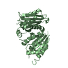
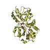
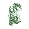
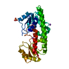
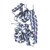
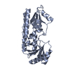
| タイトル | Crystal structure of iAChSnFR Fluorescent Acetylcholine Sensor precursor binding protein |
|---|
 要素 要素 | iAChSnFR Fluorescent Acetylcholine Sensor V9 precursor binding protein |
|---|
 キーワード キーワード | Neurotransmitter-binding protein |
|---|
| 機能・相同性 | ABC-type glycine betaine transport system, substrate-binding domain / Substrate binding domain of ABC-type glycine betaine transport system / transmembrane transporter activity / ATP-binding cassette (ABC) transporter complex / Prokaryotic membrane lipoprotein lipid attachment site profile. / ACETYLCHOLINE / DI(HYDROXYETHYL)ETHER / SPERMIDINE / Glycine/betaine ABC transporter substrate-binding protein 機能・相同性情報 機能・相同性情報 |
|---|
| 生物種 |   Thermoanaerobacter sp. X513 (バクテリア) Thermoanaerobacter sp. X513 (バクテリア) |
|---|
| 手法 |  X線回折 / シンクロトロン / 分子置換 / 解像度: 1.64 Å X線回折 / シンクロトロン / 分子置換 / 解像度: 1.64 Å |
|---|
 データ登録者 データ登録者 | Fan, C. / Borden, P.M. / Looger, L.L. / Lester, H.A. / Rees, D.C. |
|---|
| 資金援助 |  米国, 6件 米国, 6件 | 組織 | 認可番号 | 国 |
|---|
| Howard Hughes Medical Institute (HHMI) | | 米国 | | National Institutes of Health/National Institute on Drug Abuse (NIH/NIDA) | DA037161, DA043829, GM123582 | 米国 | | National Institutes of Health/National Institute on Drug Abuse (NIH/NIDA) | | | | National Institutes of Health/National Institute on Drug Abuse (NIH/NIDA) | | | | Tobacco-Related Disease Research Program (TRDRP) | 23XT-0007 | 米国 | | Tobacco-Related Disease Research Program (TRDRP) | 27IP-0057 | 米国 |
|
|---|
 引用 引用 | ジャーナル: To Be Published
タイトル: A genetically encoded fluorescent sensor for in vivo acetylcholine detection
著者: Borden, P.M. / Shivange, A.V. / Fan, C. / Rees, D.C. / Lester, H.A. / Looger, L.L. |
|---|
| 履歴 | | 登録 | 2019年11月21日 | 登録サイト: RCSB / 処理サイト: RCSB |
|---|
| 改定 1.0 | 2020年11月25日 | Provider: repository / タイプ: Initial release |
|---|
| 改定 1.1 | 2023年10月11日 | Group: Data collection / Database references / Refinement description
カテゴリ: chem_comp_atom / chem_comp_bond ...chem_comp_atom / chem_comp_bond / database_2 / pdbx_initial_refinement_model
Item: _database_2.pdbx_DOI / _database_2.pdbx_database_accession |
|---|
|
|---|
 ムービー
ムービー コントローラー


 データを開く
 基本情報
基本情報 構造の表示
構造の表示 ダウンロードとリンク
 その他のダウンロード


 PDBj
PDBj


 集合体
集合体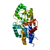
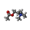


 分子量: 146.207 Da / 分子数: 1 / 由来タイプ: 合成 / 式: C7H16NO2 / タイプ: SUBJECT OF INVESTIGATION / コメント: 神経伝達物質*YM
 分子量: 207.290 Da / 分子数: 1 / 由来タイプ: 合成 / 式: C8H17NO3S / コメント: pH緩衝剤*YM
 分子量: 145.246 Da / 分子数: 1 / 由来タイプ: 合成 / 式: C7H19N3
 分子量: 106.120 Da / 分子数: 1 / 由来タイプ: 合成 / 式: C4H10O3
 試料調製
試料調製 解析
解析