 Movie
Movie Controller

[English] 日本語
 Yorodumi
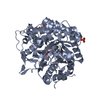
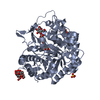
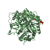
- PDB-3wq6: beta-Primeverosidase in complex with disaccharide substrate-analo... -
+ Open data

- Basic information
| Entry | Database: PDB / ID: 3wq6 | |||||||||
|---|---|---|---|---|---|---|---|---|---|---|
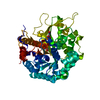
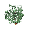
| Title | beta-Primeverosidase in complex with disaccharide substrate-analog N-beta-primeverosylamidine, artificial aglycone derivative | |||||||||
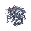
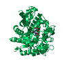
 Components Components | beta-primeverosidase | |||||||||
 Keywords Keywords | HYDROLASE / DIGLYCOSIDASE / DIGLYCOSIDE / DISACCHARIDE / GLYCOSIDE HYDROLASE FAMILY 1 (GH1) / (BETA/ALPHA)8 BARREL / SPECIFIC HYDROLYSIS OF BETA-PRIMEVEROSIDES / AROMA FORMATION / OOLONG TEA / BLACK TEA | |||||||||
| Function / homology |  Function and homology information Function and homology informationbeta-primeverosidase / beta-primeverosidase activity / beta-glucosidase activity / carbohydrate metabolic process Similarity search - Function | |||||||||
| Biological species |  Camellia sinensis (black tea) Camellia sinensis (black tea) | |||||||||
| Method |  X-RAY DIFFRACTION / MOLECULAR REPLACEMENT / Resolution: 1.8 Å X-RAY DIFFRACTION / MOLECULAR REPLACEMENT / Resolution: 1.8 Å | |||||||||
 Authors Authors | Saino, H. | |||||||||
 Citation Citation | Journal: J.Biol.Chem. / Year: 2014 Title: Crystal structures of beta-primeverosidase in complex with disaccharide amidine inhibitors. Authors: Saino, H. / Shimizu, T. / Hiratake, J. / Nakatsu, T. / Kato, H. / Sakata, K. / Mizutani, M. | |||||||||
| History |
|
- Structure visualization
| Structure viewer | Molecule: MolmilJmol/JSmol |
|---|
- Downloads & links
-Download
| PDBx/mmCIF format | 3wq6.cif.gz | 252 KB | Display | PDBx/mmCIF format |
|---|---|---|---|---|
| PDB format | pdb3wq6.ent.gz | 194.2 KB | Display | PDB format |
| PDBx/mmJSON format | 3wq6.json.gz | Tree view | PDBx/mmJSON format | |
| Others |  Other downloads Other downloads |
-Validation report
| Arichive directory | https://data.pdbj.org/pub/pdb/validation_reports/wq/3wq6ftp://data.pdbj.org/pub/pdb/validation_reports/wq/3wq6 | HTTPS FTP |
|---|
-Related structure data
| Related structure data |  3wq4C  3wq5C  1cbgS C: citing same article ( S: Starting model for refinement |
|---|---|
| Similar structure data |

-Links
 PDBj
PDBj- Assembly
| Deposited unit | 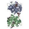
| ||||||||
|---|---|---|---|---|---|---|---|---|---|
| 1 | 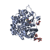
| ||||||||
| 2 | 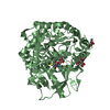
| ||||||||
| Unit cell |
|
-Components
-Protein , 1 types, 2 molecules AB
| #1: Protein | Mass: 57104.941 Da / Num. of mol.: 2 Source method: isolated from a genetically manipulated source Source: (gene. exp.) Camellia sinensis (black tea) / Production host:  Trichoplusia ni (cabbage looper) / Strain (production host): BTI-TN-5B1-4 / References: UniProt: Q7X9A9, beta-primeverosidase Trichoplusia ni (cabbage looper) / Strain (production host): BTI-TN-5B1-4 / References: UniProt: Q7X9A9, beta-primeverosidase |
|---|
-Sugars , 3 types, 4 molecules 
| #2: Polysaccharide | beta-D-mannopyranose-(1-4)-2-acetamido-2-deoxy-beta-D-glucopyranose-(1-4)-[alpha-L-fucopyranose-(1- ...beta-D-mannopyranose-(1-4)-2-acetamido-2-deoxy-beta-D-glucopyranose-(1-4)-[alpha-L-fucopyranose-(1-6)]2-acetamido-2-deoxy-beta-D-glucopyranose Source method: isolated from a genetically manipulated source |
|---|---|
| #3: Polysaccharide | 2-acetamido-2-deoxy-beta-D-glucopyranose-(1-4)-2-acetamido-2-deoxy-beta-D-glucopyranose Source method: isolated from a genetically manipulated source |
| #5: Sugar |  Type: D-saccharide, beta linking / Mass: 221.208 Da / Num. of mol.: 2 Type: D-saccharide, beta linking / Mass: 221.208 Da / Num. of mol.: 2Source method: isolated from a genetically manipulated source Formula: C8H15NO6 |
-Non-polymers , 2 types, 1783 molecules 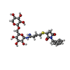

| #4: Chemical |  Mass: 599.650 Da / Num. of mol.: 2 / Source method: obtained synthetically / Formula: C26H37N3O11S Mass: 599.650 Da / Num. of mol.: 2 / Source method: obtained synthetically / Formula: C26H37N3O11S#6: Water | ChemComp-HOH / | Mass: 18.015 Da / Num. of mol.: 1781 / Source method: isolated from a natural source / Formula: H2O |
|---|
-Details
| Has protein modification | Y |
|---|
-Experimental details
-Experiment
| Experiment | Method: X-RAY DIFFRACTION / Number of used crystals: 1 |
|---|
- Sample preparation
| Crystal | Density Matthews: 2.27 Å3/Da / Density % sol: 45.93 % |
|---|---|
| Crystal grow | Temperature: 293 K / Method: vapor diffusion, hanging drop / pH: 5.5 Details: 10mg/mL protein, 0.01% TritonX-100, 21% PEG 4000, 0.3M ammonium acetate, 0.1M sodium citrate (pH 5.5), VAPOR DIFFUSION, HANGING DROP, temperature 293K |
-Data collection
| Diffraction | Mean temperature: 100 K |
|---|---|
| Diffraction source | Source: ROTATING ANODE / Type: RIGAKU FR-E SUPERBRIGHT / Wavelength: 1.5418 Å |
| Detector | Type: RIGAKU RAXIS IV / Detector: IMAGE PLATE / Date: Jun 15, 2006 |
| Radiation | Monochromator: Confocal, Multilayer Mirror / Protocol: SINGLE WAVELENGTH / Monochromatic (M) / Laue (L): M / Scattering type: x-ray |
| Radiation wavelength | Wavelength: 1.5418 Å / Relative weight: 1 |
| Reflection | Resolution: 1.8→50 Å / Num. obs: 97485 / % possible obs: 100 % / Redundancy: 3.6 % / Rmerge(I) obs: 0.068 |
| Reflection shell | Resolution: 1.8→1.9 Å / Redundancy: 3.5 % / Rmerge(I) obs: 0.257 / Mean I/σ(I) obs: 4.6 / Num. unique all: 14038 / % possible all: 100 |
- Processing
| Software |
| |||||||||||||||||||||||||||||||||||||||||||||||||||||||
|---|---|---|---|---|---|---|---|---|---|---|---|---|---|---|---|---|---|---|---|---|---|---|---|---|---|---|---|---|---|---|---|---|---|---|---|---|---|---|---|---|---|---|---|---|---|---|---|---|---|---|---|---|---|---|---|---|
| Refinement | Method to determine structure: MOLECULAR REPLACEMENT Starting model: 1CBG Resolution: 1.8→39.37 Å / SU ML: 0.16 / σ(F): 1.35 / Phase error: 15.75 / Stereochemistry target values: ML
| |||||||||||||||||||||||||||||||||||||||||||||||||||||||
| Solvent computation | Shrinkage radii: 0.9 Å / VDW probe radii: 1.11 Å / Solvent model: FLAT BULK SOLVENT MODEL | |||||||||||||||||||||||||||||||||||||||||||||||||||||||
| Refinement step | Cycle: LAST / Resolution: 1.8→39.37 Å
| |||||||||||||||||||||||||||||||||||||||||||||||||||||||
| Refine LS restraints |
| |||||||||||||||||||||||||||||||||||||||||||||||||||||||
| LS refinement shell | Refine-ID: X-RAY DIFFRACTION / Total num. of bins used: 10 / % reflection obs: 100 %
|
