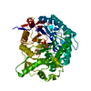
Deposited unit
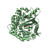
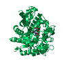
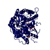
AAA: Beta-glucosidase
BBB: Beta-glucosidase
CCC: Beta-glucosidase
DDD: Beta-glucosidase
EEE: Beta-glucosidase
FFF: Beta-glucosidase
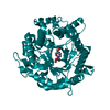
GGG: Beta-glucosidase
HHH: Beta-glucosidase
hetero molecules Summary Component details
Theoretical mass Number of molelcules Total (without water) 439,786 32 Polymers 436,782 8 Non-polymers 3,004 24 Water 35,165 1952
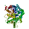
1
AAA: Beta-glucosidase
hetero molecules Summary Component details Symmetry operations
Theoretical mass Number of molelcules Total (without water) 54,965 4 Polymers 54,598 1 Non-polymers 367 3 Water 18 1
Type Name Symmetry operation Number identity operation 1_555 x,y,z 1
2
BBB: Beta-glucosidase
hetero molecules Summary Component details Symmetry operations
Theoretical mass Number of molelcules Total (without water) 55,066 5 Polymers 54,598 1 Non-polymers 469 4 Water 18 1
Type Name Symmetry operation Number identity operation 1_555 x,y,z 1
3
CCC: Beta-glucosidase
hetero molecules Summary Component details Symmetry operations
Theoretical mass Number of molelcules Total (without water) 55,053 5 Polymers 54,598 1 Non-polymers 455 4 Water 18 1
Type Name Symmetry operation Number identity operation 1_555 x,y,z 1
4
DDD: Beta-glucosidase
hetero molecules Summary Component details Symmetry operations
Theoretical mass Number of molelcules Total (without water) 54,880 2 Polymers 54,598 1 Non-polymers 282 1 Water 18 1
Type Name Symmetry operation Number identity operation 1_555 x,y,z 1
5
EEE: Beta-glucosidase
hetero molecules Summary Component details Symmetry operations
Theoretical mass Number of molelcules Total (without water) 54,903 3 Polymers 54,598 1 Non-polymers 305 2 Water 18 1
Type Name Symmetry operation Number identity operation 1_555 x,y,z 1
6
FFF: Beta-glucosidase
hetero molecules Summary Component details Symmetry operations
Theoretical mass Number of molelcules Total (without water) 54,988 5 Polymers 54,598 1 Non-polymers 390 4 Water 18 1
Type Name Symmetry operation Number identity operation 1_555 x,y,z 1
7
GGG: Beta-glucosidase
hetero molecules Summary Component details Symmetry operations
Theoretical mass Number of molelcules Total (without water) 55,050 6 Polymers 54,598 1 Non-polymers 452 5 Water 18 1
Type Name Symmetry operation Number identity operation 1_555 x,y,z 1
8
HHH: Beta-glucosidase
hetero molecules Summary Component details Symmetry operations
Theoretical mass Number of molelcules Total (without water) 54,880 2 Polymers 54,598 1 Non-polymers 282 1 Water 18 1
Type Name Symmetry operation Number identity operation 1_555 x,y,z 1
Unit cell Length a, b, c (Å) 62.654, 91.644, 159.342 Angle α, β, γ (deg.) 91.616, 90.422, 89.988 Int Tables number 1 Space group name H-M P1
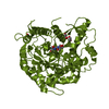
Noncrystallographic symmetry (NCS) NCS domain Show large table (3 x 28) Hide large table ID Ens-ID Details (eV)1 1 Chains A B2 1 Chains A C3 1 Chains A D4 1 Chains A E5 1 Chains A F6 1 Chains A G7 1 Chains A H8 1 Chains B C9 1 Chains B D10 1 Chains B E11 1 Chains B F12 1 Chains B G13 1 Chains B H14 1 Chains C D15 1 Chains C E16 1 Chains C F17 1 Chains C G18 1 Chains C H19 1 Chains D E20 1 Chains D F21 1 Chains D G22 1 Chains D H23 1 Chains E F24 1 Chains E G25 1 Chains E H26 1 Chains F G27 1 Chains F H28 1 Chains G H
 Movie
Movie Controller

 Yorodumi
 Open data
 Basic information
 Components
 Keywords
 Function and homology information
 Alicyclobacillus tengchongensis (bacteria)
 X-RAY DIFFRACTION /
  Authors
 Citation
 Structure visualization
 Downloads & links
 Other downloads
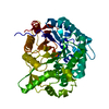


 PDBj
PDBj Assembly


 Mass: 282.334 Da / Num. of mol.: 8 / Source method: obtained synthetically / Formula: C11H26N2O6 / Comment: pH buffer*YM
 Mass: 62.068 Da / Num. of mol.: 7 / Source method: obtained synthetically / Formula: C2H6O2
 Mass: 22.990 Da / Num. of mol.: 8 / Source method: obtained synthetically / Formula: Na
 Mass: 126.904 Da / Num. of mol.: 1 / Source method: obtained synthetically / Formula: I
 Sample preparation
 / Beamline: 11.2C / Wavelength: 0.9139 Å
 Processing