 Movie
Movie Controller

[English] 日本語
 Yorodumi
- PDB-2wc3: Structure of family 1 beta-glucosidase from Thermotoga maritima i... -
+ Open data

- Basic information
| Entry | Database: PDB / ID: 2wc3 | ||||||
|---|---|---|---|---|---|---|---|
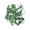
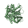
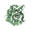
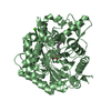
| Title | Structure of family 1 beta-glucosidase from Thermotoga maritima in complex with 3-imino-2-oxa-(+)-8-epi-castanospermine | ||||||
 Components Components | BETA-GLUCOSIDASE A | ||||||
 Keywords Keywords | HYDROLASE / CASTANOSPERMINE / GLYCOSIDE HYDROLASE / POLYSACCHARIDE DEGRADATION / CELLULOSE DEGRADATION / CARBOHYDRATE METABOLISM / FAMILY 1 / INHIBITORS / GLYCOSIDASE | ||||||
| Function / homology |  Function and homology information Function and homology informationbeta-glucosidase / beta-glucosidase activity / cellulose catabolic process Similarity search - Function | ||||||
| Biological species |   THERMOTOGA MARITIMA (bacteria) THERMOTOGA MARITIMA (bacteria) | ||||||
| Method |  X-RAY DIFFRACTION / MOLECULAR REPLACEMENT / Resolution: 2 Å X-RAY DIFFRACTION / MOLECULAR REPLACEMENT / Resolution: 2 Å | ||||||
 Authors Authors | Aguilar, M. / Gloster, T.M. / Turkenburg, J.P. / Garcia-Moreno, M.I. / Ortiz Mellet, C. / Davies, G.J. / Garcia Fernandez, J.M. | ||||||
 Citation Citation | Journal: Org.Biomol.Chem. / Year: 2009 Title: Glycosidase Inhibition by Ring-Modified Castanospermine Analogues: Tackling Enzyme Selectivity by Inhibitor Tailoring. Authors: Aguilar-Moncayo, M. / Gloster, T.M. / Turkenburg, J.P. / Garcia-Moreno, M.I. / Ortiz Mellet, C. / Davies, G.J. / Garcia Fernandez, J.M. | ||||||
| History |
| ||||||
| Remark 700 | SHEET DETERMINATION METHOD: DSSP THE SHEETS PRESENTED AS "AB" IN EACH CHAIN ON SHEET RECORDS BELOW ... SHEET DETERMINATION METHOD: DSSP THE SHEETS PRESENTED AS "AB" IN EACH CHAIN ON SHEET RECORDS BELOW IS ACTUALLY AN 8-STRANDED BARREL THIS IS REPRESENTED BY A 9-STRANDED SHEET IN WHICH THE FIRST AND LAST STRANDS ARE IDENTICAL. THE SHEETS PRESENTED AS "BB" IN EACH CHAIN ON SHEET RECORDS BELOW IS ACTUALLY AN 8-STRANDED BARREL THIS IS REPRESENTED BY A 9-STRANDED SHEET IN WHICH THE FIRST AND LAST STRANDS ARE IDENTICAL. THE SHEETS PRESENTED AS "CB" IN EACH CHAIN ON SHEET RECORDS BELOW IS ACTUALLY AN 9-STRANDED BARREL THIS IS REPRESENTED BY A 10-STRANDED SHEET IN WHICH THE FIRST AND LAST STRANDS ARE IDENTICAL. THE SHEETS PRESENTED AS "DB" IN EACH CHAIN ON SHEET RECORDS BELOW IS ACTUALLY AN 7-STRANDED BARREL THIS IS REPRESENTED BY A 8-STRANDED SHEET IN WHICH THE FIRST AND LAST STRANDS ARE IDENTICAL. |
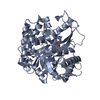
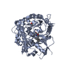
- Structure visualization
| Structure viewer | Molecule: MolmilJmol/JSmol |
|---|
- Downloads & links
-Download
| PDBx/mmCIF format | 2wc3.cif.gz | 385.5 KB | Display | PDBx/mmCIF format |
|---|---|---|---|---|
| PDB format | pdb2wc3.ent.gz | 315.4 KB | Display | PDB format |
| PDBx/mmJSON format | 2wc3.json.gz | Tree view | PDBx/mmJSON format | |
| Others |  Other downloads Other downloads |
-Validation report
| Arichive directory | https://data.pdbj.org/pub/pdb/validation_reports/wc/2wc3ftp://data.pdbj.org/pub/pdb/validation_reports/wc/2wc3 | HTTPS FTP |
|---|
-Related structure data
| Related structure data | 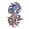 2wbgC 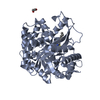 2wc4C  1od0S C: citing same article ( S: Starting model for refinement |
|---|---|
| Similar structure data |


-Links
 PDBj
PDBj- Assembly
| Deposited unit | 
| ||||||||
|---|---|---|---|---|---|---|---|---|---|
| 1 | 
| ||||||||
| 2 | 
| ||||||||
| 3 | 
| ||||||||
| 4 | 
| ||||||||
| Unit cell |
|
-Components
| #1: Protein | Mass: 53940.648 Da / Num. of mol.: 4 / Fragment: RESIDUES 2-446 Source method: isolated from a genetically manipulated source Source: (gene. exp.) THERMOTOGA MARITIMA (bacteria) / Production host: #2: Chemical | ChemComp-AM3 / ( 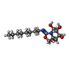  Mass: 316.393 Da / Num. of mol.: 4 / Source method: obtained synthetically / Formula: C15H28N2O5 Mass: 316.393 Da / Num. of mol.: 4 / Source method: obtained synthetically / Formula: C15H28N2O5#3: Water | ChemComp-HOH / |  Mass: 18.015 Da / Num. of mol.: 1118 / Source method: isolated from a natural source / Formula: H2O Mass: 18.015 Da / Num. of mol.: 1118 / Source method: isolated from a natural source / Formula: H2OSequence details | THE SEQUENCE CRYSTALLIZED CONTAINS AN N-TERMINAL HIS TAG FROM THE EXPRESSION VECTOR. NUMBERING ...THE SEQUENCE CRYSTALLIZ | |
|---|
-Experimental details
-Experiment
| Experiment | Method: X-RAY DIFFRACTION / Number of used crystals: 1 |
|---|
- Sample preparation
| Crystal | Density Matthews: 2 Å3/Da / Density % sol: 39 % / Description: NONE |
|---|---|
| Crystal grow | pH: 7 Details: 10 MG/ML PROTEIN, 0.1 M IMIDAZOLE, PH7, 0.2 CALCIUM ACETATE, 15-17% PEG 4000 |
-Data collection
| Diffraction | Mean temperature: 120 K |
|---|---|
| Diffraction source | Source: ROTATING ANODE / Type: RIGAKU MICROMAX-007 HF / Wavelength: 1.5418 |
| Radiation | Protocol: SINGLE WAVELENGTH / Monochromatic (M) / Laue (L): M / Scattering type: x-ray |
| Radiation wavelength | Wavelength: 1.5418 Å / Relative weight: 1 |
| Reflection | Resolution: 2→25 Å / Num. obs: 112653 / % possible obs: 97.1 % / Redundancy: 3.1 % / Rmerge(I) obs: 0.11 / Net I/σ(I): 6.5 |
| Reflection shell | Resolution: 2→2.07 Å / Redundancy: 3.1 % / Rmerge(I) obs: 0.36 / Mean I/σ(I) obs: 2.4 / % possible all: 90.7 |
- Processing
| Software |
| ||||||||||||||||||||||||||||||||||||||||||||||||||||||||||||||||||||||||||||||||||||||||||||||||||||||||||||||||||||||||||||||||||||||||||||||||||||||||||||||||||||||||||||||||||||||
|---|---|---|---|---|---|---|---|---|---|---|---|---|---|---|---|---|---|---|---|---|---|---|---|---|---|---|---|---|---|---|---|---|---|---|---|---|---|---|---|---|---|---|---|---|---|---|---|---|---|---|---|---|---|---|---|---|---|---|---|---|---|---|---|---|---|---|---|---|---|---|---|---|---|---|---|---|---|---|---|---|---|---|---|---|---|---|---|---|---|---|---|---|---|---|---|---|---|---|---|---|---|---|---|---|---|---|---|---|---|---|---|---|---|---|---|---|---|---|---|---|---|---|---|---|---|---|---|---|---|---|---|---|---|---|---|---|---|---|---|---|---|---|---|---|---|---|---|---|---|---|---|---|---|---|---|---|---|---|---|---|---|---|---|---|---|---|---|---|---|---|---|---|---|---|---|---|---|---|---|---|---|---|---|
| Refinement | Method to determine structure: MOLECULAR REPLACEMENT Starting model: PDB ENTRY 1OD0 Resolution: 2→113.23 Å / Cor.coef. Fo:Fc: 0.957 / Cor.coef. Fo:Fc free: 0.919 / SU B: 4.893 / SU ML: 0.137 / Cross valid method: THROUGHOUT / ESU R: 0.228 / ESU R Free: 0.197 / Stereochemistry target values: MAXIMUM LIKELIHOOD / Details: HYDROGENS HAVE BEEN ADDED IN THE RIDING POSITIONS.
| ||||||||||||||||||||||||||||||||||||||||||||||||||||||||||||||||||||||||||||||||||||||||||||||||||||||||||||||||||||||||||||||||||||||||||||||||||||||||||||||||||||||||||||||||||||||
| Solvent computation | Ion probe radii: 0.8 Å / Shrinkage radii: 0.8 Å / VDW probe radii: 1.2 Å / Solvent model: MASK | ||||||||||||||||||||||||||||||||||||||||||||||||||||||||||||||||||||||||||||||||||||||||||||||||||||||||||||||||||||||||||||||||||||||||||||||||||||||||||||||||||||||||||||||||||||||
| Displacement parameters | Biso mean: 26.91 Å2
| ||||||||||||||||||||||||||||||||||||||||||||||||||||||||||||||||||||||||||||||||||||||||||||||||||||||||||||||||||||||||||||||||||||||||||||||||||||||||||||||||||||||||||||||||||||||
| Refinement step | Cycle: LAST / Resolution: 2→113.23 Å
| ||||||||||||||||||||||||||||||||||||||||||||||||||||||||||||||||||||||||||||||||||||||||||||||||||||||||||||||||||||||||||||||||||||||||||||||||||||||||||||||||||||||||||||||||||||||
| Refine LS restraints |
|