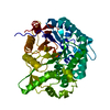
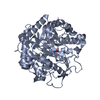
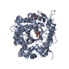
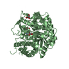
Glycoside hydrolase, family 1, beta-glucosidase / Glycoside hydrolase family 1, active site / Glycosyl hydrolases family 1 active site. / Glycosyl hydrolases family 1, N-terminal conserved site / Glycosyl hydrolases family 1 N-terminal signature. / Glycosyl hydrolase family 1 / Glycoside hydrolase family 1 / Glycosidases / Glycoside hydrolase superfamily / TIM Barrel ...Glycoside hydrolase, family 1, beta-glucosidase / Glycoside hydrolase family 1, active site / Glycosyl hydrolases family 1 active site. / Glycosyl hydrolases family 1, N-terminal conserved site / Glycosyl hydrolases family 1 N-terminal signature. / Glycosyl hydrolase family 1 / Glycoside hydrolase family 1 / Glycosidases / Glycoside hydrolase superfamily / TIM Barrel / Alpha-Beta Barrel / Alpha Beta Similarity search - Domain/homology
Resolution: 2.1→56.41 Å / Cor.coef. Fo:Fc: 0.959 / Cor.coef. Fo:Fc free: 0.932 / SU B: 5.78 / SU ML: 0.149 / Cross valid method: THROUGHOUT / ESU R: 0.207 / ESU R Free: 0.183 / Stereochemistry target values: MAXIMUM LIKELIHOOD / Details: HYDROGENS HAVE BEEN ADDED IN THE RIDING POSITIONS
Rfactor
Num. reflection
% reflection
Selection details
Rfree
0.23666
3042
5.1 %
RANDOM
Rwork
0.18396
-
-
-
obs
0.18671
57056
99.99 %
-
Solvent computation
Ion probe radii: 0.8 Å / Shrinkage radii: 0.8 Å / VDW probe radii: 1.2 Å / Solvent model: MASK
 Movie
Movie Controller

 Yorodumi
 Open data
 Basic information
 Components
 Keywords
 Function and homology information
 Thermotoga maritima (bacteria)
 X-RAY DIFFRACTION /
  Authors
 United Kingdom, 1items
  Citation
 Structure visualization
 Downloads & links
 Other downloads


 PDBj
PDBj Assembly


 Mass: 35.453 Da / Num. of mol.: 1 / Source method: obtained synthetically / Formula: Cl
 Mass: 62.068 Da / Num. of mol.: 6 / Source method: obtained synthetically / Formula: C2H6O2
 Mass: 187.173 Da / Num. of mol.: 2 / Source method: obtained synthetically / Formula: C7H11N2O4
 Mass: 18.015 Da / Num. of mol.: 262 / Source method: isolated from a natural source / Formula: H2O
 Sample preparation
 / Beamline: ID23-2 / Wavelength: 0.8726 Å
 Processing