 Movie
Movie Controller


+ Open data

- Basic information
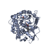
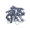
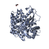
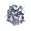
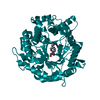
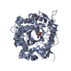
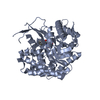
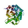
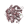
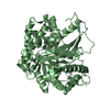
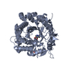
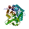
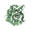
| Entry | Database: PDB / ID: 1oin | ||||||
|---|---|---|---|---|---|---|---|
| Title | Family 1 b-glucosidase from Thermotoga maritima | ||||||
 Components Components | BETA-GLUCOSIDASE A | ||||||
 Keywords Keywords | HYDROLASE / GLUCOSIDE HYDROLYSIS / FAMILY GH1 | ||||||
| Function / homology |  Function and homology information Function and homology informationbeta-glucosidase / beta-glucosidase activity / cellulose catabolic process Similarity search - Function | ||||||
| Biological species |   THERMOTOGA MARITIMA (bacteria) THERMOTOGA MARITIMA (bacteria) | ||||||
| Method |  X-RAY DIFFRACTION / SYNCHROTRON / MOLECULAR REPLACEMENT / Resolution: 2.15 Å X-RAY DIFFRACTION / SYNCHROTRON / MOLECULAR REPLACEMENT / Resolution: 2.15 Å | ||||||
 Authors Authors | Gloster, T. / Zechel, D.L. / Boraston, A.B. / Boraston, C.M. / Macdonald, J.M. / Tilbrook, D.M. / Stick, R.V. / Davies, G.J. | ||||||
 Citation Citation | Journal: J.Am.Chem.Soc. / Year: 2003 Title: Iminosugar Glycosidase Inhibitors: Structural and Thermodynamic Dissection of the Binding of Isofagomine and 1-Deoxynojirimycin to Beta-Glucosidases Authors: Zechel, D.L. / Boraston, A.B. / Gloster, T. / Boraston, C.M. / Macdonald, J.M. / Tilbrook, D.M. / Stick, R.V. / Davies, G.J. | ||||||
| History |
| ||||||
| Remark 700 | SHEET DETERMINATION METHOD: DSSP THE SHEETS PRESENTED AS "AB" IN EACH CHAIN ON SHEET RECORDS BELOW ... SHEET DETERMINATION METHOD: DSSP THE SHEETS PRESENTED AS "AB" IN EACH CHAIN ON SHEET RECORDS BELOW IS ACTUALLY AN 8-STRANDED BARREL THIS IS REPRESENTED BY A 9-STRANDED SHEET IN WHICH THE FIRST AND LAST STRANDS ARE IDENTICAL. THE SHEETS PRESENTED AS "BB" IN EACH CHAIN ON SHEET RECORDS BELOW IS ACTUALLY AN 8-STRANDED BARREL THIS IS REPRESENTED BY A 9-STRANDED SHEET IN WHICH THE FIRST AND LAST STRANDS ARE IDENTICAL. |
- Structure visualization
| Structure viewer | Molecule: MolmilJmol/JSmol |
|---|
- Downloads & links
-Download
| PDBx/mmCIF format | 1oin.cif.gz | 197.7 KB | Display | PDBx/mmCIF format |
|---|---|---|---|---|
| PDB format | pdb1oin.ent.gz | 156 KB | Display | PDB format |
| PDBx/mmJSON format | 1oin.json.gz | Tree view | PDBx/mmJSON format | |
| Others |  Other downloads Other downloads |
-Validation report
| Arichive directory | https://data.pdbj.org/pub/pdb/validation_reports/oi/1oinftp://data.pdbj.org/pub/pdb/validation_reports/oi/1oin | HTTPS FTP |
|---|
-Related structure data
| Related structure data |  1od0SC 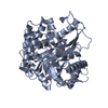 1oifC  1oimC C: citing same article ( S: Starting model for refinement |
|---|---|
| Similar structure data |


-Links
 PDBj
PDBj- Assembly
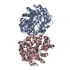
| Deposited unit | 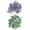
| |||||||||||||||||||||||||||
|---|---|---|---|---|---|---|---|---|---|---|---|---|---|---|---|---|---|---|---|---|---|---|---|---|---|---|---|---|
| 1 | 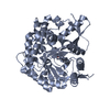
| |||||||||||||||||||||||||||
| 2 | 
| |||||||||||||||||||||||||||
| Unit cell |
| |||||||||||||||||||||||||||
| Noncrystallographic symmetry (NCS) | NCS domain:
NCS domain segments:
NCS oper: (Code: given Matrix: (0.99952, -0.02596, 0.01718), Vector: |
-Components
| #1: Protein | Mass: 53940.648 Da / Num. of mol.: 2 Source method: isolated from a genetically manipulated source Source: (gene. exp.) THERMOTOGA MARITIMA (bacteria) / Production host: #2: Sugar |   Type: D-saccharide, alpha linking / Mass: 182.147 Da / Num. of mol.: 2 Type: D-saccharide, alpha linking / Mass: 182.147 Da / Num. of mol.: 2Source method: isolated from a genetically manipulated source Formula: C6H11FO5 #3: Water | ChemComp-HOH / |  Mass: 18.015 Da / Num. of mol.: 413 / Source method: isolated from a natural source / Formula: H2O Mass: 18.015 Da / Num. of mol.: 413 / Source method: isolated from a natural source / Formula: H2OCompound details | CATALYTIC ACTIVITY: RELEASE OF BETA-D-GLUCOSE DURING HYDROLYSIS OF TERMINAL, NON-REDUCING BETA-D- ...CATALYTIC ACTIVITY: RELEASE OF BETA-D-GLUCOSE DURING HYDROLYSIS | Has protein modification | Y | Sequence details | A1,305-306, 446 B1,213,446 POOR OR MISSING DENSITY THE FULL LENGTH PROTEIN ALSO CONTAINS A HIS-TAG ...A1,305-306, 446 B1,213,446 POOR OR MISSING DENSITY THE FULL LENGTH PROTEIN ALSO CONTAINS A HIS-TAG FROM THE PET28A CLONING VECTOR. NUMBERING OF THE PROTEIN AGREES WITH THE SWISSPROT ENTRY AND THUS IGNORES THE HIS-TAG | |
|---|
-Experimental details
-Experiment
| Experiment | Method: X-RAY DIFFRACTION / Number of used crystals: 1 |
|---|
- Sample preparation
| Crystal | Density Matthews: 2.3 Å3/Da / Density % sol: 46 % / Description: STRUCTURE ISOMORPHOUS WITH STARTING ENTRY | ||||||||||||||||||||||||||||||||||||||||||
|---|---|---|---|---|---|---|---|---|---|---|---|---|---|---|---|---|---|---|---|---|---|---|---|---|---|---|---|---|---|---|---|---|---|---|---|---|---|---|---|---|---|---|---|
| Crystal grow | pH: 7 Details: 10MG/ML PROTEIN IN 15% PEG 4K, 0.2 M CAAC, 0.1M IMIDAZOLE PH7, pH 7.00 | ||||||||||||||||||||||||||||||||||||||||||
| Crystal grow | *PLUS Temperature: 18 ℃ / pH: 7 / Method: vapor diffusion, hanging drop | ||||||||||||||||||||||||||||||||||||||||||
| Components of the solutions | *PLUS
|
-Data collection
| Diffraction | Mean temperature: 100 K |
|---|---|
| Diffraction source | Source: SYNCHROTRON / Site: ESRF  / Beamline: ID14-2 / Wavelength: 0.933 / Beamline: ID14-2 / Wavelength: 0.933 |
| Detector | Type: ADSC CCD / Detector: CCD / Date: Dec 15, 2001 |
| Radiation | Protocol: SINGLE WAVELENGTH / Monochromatic (M) / Laue (L): M / Scattering type: x-ray |
| Radiation wavelength | Wavelength: 0.933 Å / Relative weight: 1 |
| Reflection | Resolution: 2.15→25 Å / Num. obs: 56583 / % possible obs: 100 % / Redundancy: 5.4 % / Rmerge(I) obs: 0.055 / Net I/σ(I): 25.56 |
| Reflection shell | Resolution: 2.15→2.23 Å / Redundancy: 5.4 % / Rmerge(I) obs: 0.331 / Mean I/σ(I) obs: 5.14 / % possible all: 100 |
| Reflection | *PLUS Highest resolution: 2.15 Å / Lowest resolution: 20 Å / % possible obs: 100 % / Redundancy: 5.4 % / Rmerge(I) obs: 0.057 |
| Reflection shell | *PLUS Lowest resolution: 2.2 Å / % possible obs: 100 % / Redundancy: 5.4 % / Rmerge(I) obs: 0.4 / Mean I/σ(I) obs: 5.1 |
- Processing
| Software |
| ||||||||||||||||||||||||||||||||||||||||||||||||||||||||||||||||||||||||||||||||||||||||||||||||||||||||||||||||||||||||||||||||||||||||||||||||||||||||||||||||||||||||||||||||||||||
|---|---|---|---|---|---|---|---|---|---|---|---|---|---|---|---|---|---|---|---|---|---|---|---|---|---|---|---|---|---|---|---|---|---|---|---|---|---|---|---|---|---|---|---|---|---|---|---|---|---|---|---|---|---|---|---|---|---|---|---|---|---|---|---|---|---|---|---|---|---|---|---|---|---|---|---|---|---|---|---|---|---|---|---|---|---|---|---|---|---|---|---|---|---|---|---|---|---|---|---|---|---|---|---|---|---|---|---|---|---|---|---|---|---|---|---|---|---|---|---|---|---|---|---|---|---|---|---|---|---|---|---|---|---|---|---|---|---|---|---|---|---|---|---|---|---|---|---|---|---|---|---|---|---|---|---|---|---|---|---|---|---|---|---|---|---|---|---|---|---|---|---|---|---|---|---|---|---|---|---|---|---|---|---|
| Refinement | Method to determine structure: MOLECULAR REPLACEMENT Starting model: PDB ENTRY 1OD0 Resolution: 2.15→72.55 Å / Cor.coef. Fo:Fc: 0.954 / Cor.coef. Fo:Fc free: 0.925 / SU B: 5.705 / SU ML: 0.148 / Cross valid method: THROUGHOUT / ESU R: 0.249 / ESU R Free: 0.21 / Stereochemistry target values: MAXIMUM LIKELIHOOD
| ||||||||||||||||||||||||||||||||||||||||||||||||||||||||||||||||||||||||||||||||||||||||||||||||||||||||||||||||||||||||||||||||||||||||||||||||||||||||||||||||||||||||||||||||||||||
| Solvent computation | Ion probe radii: 0.8 Å / Shrinkage radii: 0.8 Å / VDW probe radii: 1.4 Å / Solvent model: BABINET MODEL WITH MASK | ||||||||||||||||||||||||||||||||||||||||||||||||||||||||||||||||||||||||||||||||||||||||||||||||||||||||||||||||||||||||||||||||||||||||||||||||||||||||||||||||||||||||||||||||||||||
| Displacement parameters | Biso mean: 40.28 Å2
| ||||||||||||||||||||||||||||||||||||||||||||||||||||||||||||||||||||||||||||||||||||||||||||||||||||||||||||||||||||||||||||||||||||||||||||||||||||||||||||||||||||||||||||||||||||||
| Refinement step | Cycle: LAST / Resolution: 2.15→72.55 Å
| ||||||||||||||||||||||||||||||||||||||||||||||||||||||||||||||||||||||||||||||||||||||||||||||||||||||||||||||||||||||||||||||||||||||||||||||||||||||||||||||||||||||||||||||||||||||
| Refine LS restraints |
|