 Movie
Movie Controller


+ Open data

- Basic information
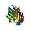
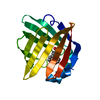
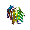
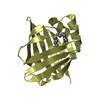
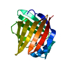
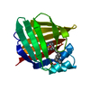
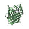
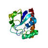
| Entry | Database: PDB / ID: 3vg2 | ||||||
|---|---|---|---|---|---|---|---|
| Title | Iodide derivative of human LFABP | ||||||
 Components Components | Fatty acid-binding protein, liver | ||||||
 Keywords Keywords | LIPID BINDING PROTEIN / LFABP / Iodide / Copper Kalpha / Palmitic acid | ||||||
| Function / homology |  Function and homology information Function and homology informationcellular detoxification / Heme degradation / Triglyceride catabolism / antioxidant activity / peroxisomal matrix / fatty acid transport / Regulation of lipid metabolism by PPARalpha / fatty acid binding / PPARA activates gene expression / Cytoprotection by HMOX1 ...cellular detoxification / Heme degradation / Triglyceride catabolism / antioxidant activity / peroxisomal matrix / fatty acid transport / Regulation of lipid metabolism by PPARalpha / fatty acid binding / PPARA activates gene expression / Cytoprotection by HMOX1 / cellular response to hydrogen peroxide / cellular response to hypoxia / chromatin binding / extracellular exosome / nucleoplasm / nucleus / cytosol Similarity search - Function | ||||||
| Biological species |  Homo Sapiens (human) Homo Sapiens (human) | ||||||
| Method |  X-RAY DIFFRACTION / MOLECULAR REPLACEMENT / molecular replacement / Resolution: 2.4 Å X-RAY DIFFRACTION / MOLECULAR REPLACEMENT / molecular replacement / Resolution: 2.4 Å | ||||||
 Authors Authors | Sharma, A. / Yogavel, M. / Sharma, A. | ||||||
 Citation Citation | Journal: J.Struct.Funct.Genom. / Year: 2012 Title: Utility of anion and cation combinations for phasing of protein structures. Authors: Sharma, A. / Yogavel, M. / Sharma, A. | ||||||
| History |
|
- Structure visualization
| Structure viewer | Molecule: MolmilJmol/JSmol |
|---|
- Downloads & links
-Download
| PDBx/mmCIF format | 3vg2.cif.gz | 40.4 KB | Display | PDBx/mmCIF format |
|---|---|---|---|---|
| PDB format | pdb3vg2.ent.gz | 27.3 KB | Display | PDB format |
| PDBx/mmJSON format | 3vg2.json.gz | Tree view | PDBx/mmJSON format | |
| Others |  Other downloads Other downloads |
-Validation report
| Arichive directory | https://data.pdbj.org/pub/pdb/validation_reports/vg/3vg2ftp://data.pdbj.org/pub/pdb/validation_reports/vg/3vg2 | HTTPS FTP |
|---|
-Related structure data
| Related structure data |  3b2hC  3b2iC  3b2jC  3b2kC  3b2lC  3vg3C  3vg4C  3vg5C  3vg6C  3vg7C C: citing same article ( |
|---|---|
| Similar structure data |

-Links
 PDBj
PDBj


- Assembly
| Deposited unit | 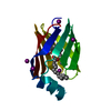
| ||||||||
|---|---|---|---|---|---|---|---|---|---|
| 1 |
| ||||||||
| Unit cell |
|
-Components
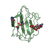
| #1: Protein | Mass: 14599.728 Da / Num. of mol.: 1 Source method: isolated from a genetically manipulated source Source: (gene. exp.) Homo Sapiens (human) / Gene: FABP1, FABPL / Plasmid: PET28a / Production host:  | ||||
|---|---|---|---|---|---|
| #2: Chemical |   Mass: 256.424 Da / Num. of mol.: 2 / Source method: obtained synthetically / Formula: C16H32O2 Mass: 256.424 Da / Num. of mol.: 2 / Source method: obtained synthetically / Formula: C16H32O2#3: Chemical | ChemComp-IOD /   Mass: 126.904 Da / Num. of mol.: 5 / Source method: obtained synthetically / Formula: I Mass: 126.904 Da / Num. of mol.: 5 / Source method: obtained synthetically / Formula: I#4: Water | ChemComp-HOH / |  Mass: 18.015 Da / Num. of mol.: 64 / Source method: isolated from a natural source / Formula: H2O Mass: 18.015 Da / Num. of mol.: 64 / Source method: isolated from a natural source / Formula: H2O |
-Experimental details
-Experiment
| Experiment | Method: X-RAY DIFFRACTION / Number of used crystals: 1 |
|---|
- Sample preparation
| Crystal | Density Matthews: 2.15 Å3/Da / Density % sol: 42.7 % / Mosaicity: 0.624 ° |
|---|---|
| Crystal grow | Temperature: 293 K / Method: hanging drop / pH: 8 Details: 30% PEG MME 2000, 0.15M KBr, pH 8.0, hanging drop, temperature 293K |
-Data collection
| Diffraction | Mean temperature: 100 K | |||||||||||||||||||||||||||||||||||||||||||||||||||||||||||||||||||||||||||||
|---|---|---|---|---|---|---|---|---|---|---|---|---|---|---|---|---|---|---|---|---|---|---|---|---|---|---|---|---|---|---|---|---|---|---|---|---|---|---|---|---|---|---|---|---|---|---|---|---|---|---|---|---|---|---|---|---|---|---|---|---|---|---|---|---|---|---|---|---|---|---|---|---|---|---|---|---|---|---|
| Diffraction source | Source: ROTATING ANODE / Type: RIGAKU MICROMAX-007 / Wavelength: 1.5418 Å | |||||||||||||||||||||||||||||||||||||||||||||||||||||||||||||||||||||||||||||
| Detector | Type: MAR scanner 345 mm plate / Detector: IMAGE PLATE / Date: Sep 10, 2009 / Details: mirrors | |||||||||||||||||||||||||||||||||||||||||||||||||||||||||||||||||||||||||||||
| Radiation | Monochromator: SAGITALLY FOCUSED Si(111) / Protocol: SINGLE WAVELENGTH / Monochromatic (M) / Laue (L): M / Scattering type: x-ray | |||||||||||||||||||||||||||||||||||||||||||||||||||||||||||||||||||||||||||||
| Radiation wavelength | Wavelength: 1.5418 Å / Relative weight: 1 | |||||||||||||||||||||||||||||||||||||||||||||||||||||||||||||||||||||||||||||
| Reflection | Resolution: 2.22→50 Å / Num. obs: 6640 / % possible obs: 99.5 % / Observed criterion σ(F): 2 / Observed criterion σ(I): 2 / Redundancy: 5.6 % / Rmerge(I) obs: 0.103 / Χ2: 1.3 / Net I/σ(I): 8 | |||||||||||||||||||||||||||||||||||||||||||||||||||||||||||||||||||||||||||||
| Reflection shell |
|
-Phasing
| Phasing | Method: molecular replacement |
|---|
- Processing
| Software |
| ||||||||||||||||||||||||||||||||||||||||||||||||||||||||||||||||||||||||||||||||||||||||||
|---|---|---|---|---|---|---|---|---|---|---|---|---|---|---|---|---|---|---|---|---|---|---|---|---|---|---|---|---|---|---|---|---|---|---|---|---|---|---|---|---|---|---|---|---|---|---|---|---|---|---|---|---|---|---|---|---|---|---|---|---|---|---|---|---|---|---|---|---|---|---|---|---|---|---|---|---|---|---|---|---|---|---|---|---|---|---|---|---|---|---|---|
| Refinement | Method to determine structure: MOLECULAR REPLACEMENT / Resolution: 2.4→8 Å / Cor.coef. Fo:Fc: 0.926 / Cor.coef. Fo:Fc free: 0.837 / WRfactor Rfree: 0.2768 / WRfactor Rwork: 0.185 / Occupancy max: 1 / Occupancy min: 0.2 / FOM work R set: 0.7832 / SU B: 10.452 / SU ML: 0.246 / SU R Cruickshank DPI: 1.2837 / SU Rfree: 0.3804 / Cross valid method: THROUGHOUT / σ(F): 0 / ESU R Free: 0.38 / Stereochemistry target values: MAXIMUM LIKELIHOOD / Details: HYDROGENS HAVE BEEN ADDED IN THE RIDING POSITIONS
| ||||||||||||||||||||||||||||||||||||||||||||||||||||||||||||||||||||||||||||||||||||||||||
| Solvent computation | Ion probe radii: 0.8 Å / Shrinkage radii: 0.8 Å / VDW probe radii: 1.2 Å / Solvent model: MASK | ||||||||||||||||||||||||||||||||||||||||||||||||||||||||||||||||||||||||||||||||||||||||||
| Displacement parameters | Biso max: 45.01 Å2 / Biso mean: 23.3338 Å2 / Biso min: 9.13 Å2
| ||||||||||||||||||||||||||||||||||||||||||||||||||||||||||||||||||||||||||||||||||||||||||
| Refinement step | Cycle: LAST / Resolution: 2.4→8 Å
| ||||||||||||||||||||||||||||||||||||||||||||||||||||||||||||||||||||||||||||||||||||||||||
| Refine LS restraints |
| ||||||||||||||||||||||||||||||||||||||||||||||||||||||||||||||||||||||||||||||||||||||||||
| LS refinement shell | Resolution: 2.4→2.457 Å / Total num. of bins used: 20
|
