+ Open data
- Basic information
| Entry | Database: PDB / ID: 3vg7 | ||||||
|---|---|---|---|---|---|---|---|
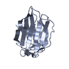
| Title | Structure of human LFABP at high resolution from S-SAD | ||||||
 Components Components | Fatty acid-binding protein, liver | ||||||
 Keywords Keywords | LIPID BINDING PROTEIN / LFABP / S-SAD / Copper Kalpha / Palmitic acid | ||||||
| Function / homology |  Function and homology information Function and homology informationcellular detoxification / Heme degradation / Triglyceride catabolism / antioxidant activity / peroxisomal matrix / fatty acid transport / Regulation of lipid metabolism by PPARalpha / fatty acid binding / PPARA activates gene expression / Cytoprotection by HMOX1 ...cellular detoxification / Heme degradation / Triglyceride catabolism / antioxidant activity / peroxisomal matrix / fatty acid transport / Regulation of lipid metabolism by PPARalpha / fatty acid binding / PPARA activates gene expression / Cytoprotection by HMOX1 / cellular response to hydrogen peroxide / cellular response to hypoxia / chromatin binding / extracellular exosome / nucleoplasm / nucleus / cytosol Similarity search - Function | ||||||
| Biological species |  Homo Sapiens (human) Homo Sapiens (human) | ||||||
| Method |  X-RAY DIFFRACTION / AB INITIO / X-RAY DIFFRACTION / AB INITIO /  SAD / Resolution: 1.44 Å SAD / Resolution: 1.44 Å | ||||||
 Authors Authors | Sharma, A. / Yogavel, M. / Sharma, A. | ||||||
 Citation Citation |  Journal: J.Struct.Funct.Genom. / Year: 2012 Journal: J.Struct.Funct.Genom. / Year: 2012Title: Utility of anion and cation combinations for phasing of protein structures. Authors: Sharma, A. / Yogavel, M. / Sharma, A. | ||||||
| History |
|
- Structure visualization
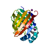
| Structure viewer | Molecule:  Molmil Molmil Jmol/JSmol Jmol/JSmol |
|---|
- Downloads & links
- Download
| PDBx/mmCIF format |  3vg7.cif.gz 3vg7.cif.gz | 46.3 KB | Display |  PDBx/mmCIF format PDBx/mmCIF format |
|---|---|---|---|---|
| PDB format |  pdb3vg7.ent.gz pdb3vg7.ent.gz | 32 KB | Display |  PDB format PDB format |
| PDBx/mmJSON format |  3vg7.json.gz 3vg7.json.gz | Tree view |  PDBx/mmJSON format PDBx/mmJSON format | |
| Others |  Other downloads Other downloads |
-Validation report
| Arichive directory |  https://data.pdbj.org/pub/pdb/validation_reports/vg/3vg7 https://data.pdbj.org/pub/pdb/validation_reports/vg/3vg7 ftp://data.pdbj.org/pub/pdb/validation_reports/vg/3vg7 ftp://data.pdbj.org/pub/pdb/validation_reports/vg/3vg7 | HTTPS FTP |
|---|
-Related structure data
| Related structure data |  3b2hC  3b2iC  3b2jC  3b2kC  3b2lC  3vg2C  3vg3C  3vg4C  3vg5C  3vg6C C: citing same article ( |
|---|---|
| Similar structure data |
- Links
Links
- Assembly
| Deposited unit | 
| ||||||||
|---|---|---|---|---|---|---|---|---|---|
| 1 |
| ||||||||
| Unit cell |
|
- Components
| #1: Protein | Mass: 14599.728 Da / Num. of mol.: 1 Source method: isolated from a genetically manipulated source Source: (gene. exp.)  Homo Sapiens (human) / Gene: FABP1, FABPL / Plasmid: PET28a / Production host: Homo Sapiens (human) / Gene: FABP1, FABPL / Plasmid: PET28a / Production host:  | ||
|---|---|---|---|
| #2: Chemical | | #3: Water | ChemComp-HOH / | |
-Experimental details
-Experiment
| Experiment | Method:  X-RAY DIFFRACTION / Number of used crystals: 1 X-RAY DIFFRACTION / Number of used crystals: 1 |
|---|
- Sample preparation
| Crystal | Density Matthews: 1.94 Å3/Da / Density % sol: 44.22 % / Description: THE FILE CONTAINS FRIEDEL PAIRS. |
|---|---|
| Crystal grow | Temperature: 293 K / Method: hanging drop / pH: 8 Details: 30% PEG MME 2000, 0.15M KBr, pH 8.0, hanging drop, temperature 293K |
-Data collection
| Diffraction | Mean temperature: 100 K |
|---|---|
| Diffraction source | Source:  ROTATING ANODE / Type: RIGAKU MICROMAX-007 / Wavelength: 1.5418 Å ROTATING ANODE / Type: RIGAKU MICROMAX-007 / Wavelength: 1.5418 Å |
| Detector | Type: MAR scanner 345 mm plate / Detector: IMAGE PLATE / Date: Sep 10, 2009 / Details: mirrors |
| Radiation | Monochromator: SAGITALLY FOCUSED Si(111) / Protocol: SINGLE WAVELENGTH / Monochromatic (M) / Laue (L): M / Scattering type: x-ray |
| Radiation wavelength | Wavelength: 1.5418 Å / Relative weight: 1 |
| Reflection | Resolution: 1.43→50 Å / Num. obs: 24038 / % possible obs: 97.3 % / Observed criterion σ(F): 2 / Observed criterion σ(I): 2 / Redundancy: 2.4 % / Rmerge(I) obs: 0.051 / Net I/σ(I): 103.7 |
| Reflection shell | Resolution: 1.43→1.48 Å / Redundancy: 23.2 % / Rmerge(I) obs: 0.417 / Mean I/σ(I) obs: 9.8 / Num. unique all: 2020 / % possible all: 84.6 |
-Phasing
| Phasing | Method:  SAD SAD |
|---|
- Processing
| Software |
| ||||||||||||||||||||||||||||||||
|---|---|---|---|---|---|---|---|---|---|---|---|---|---|---|---|---|---|---|---|---|---|---|---|---|---|---|---|---|---|---|---|---|---|
| Refinement | Method to determine structure: AB INITIO / Resolution: 1.44→6.5 Å / Num. parameters: 10853 / Num. restraintsaints: 13584 / Occupancy max: 1.15 / σ(F): 0 / Stereochemistry target values: ENGH & HUBER / Details: THE FILE CONTAINS FRIEDEL PAIRS.
| ||||||||||||||||||||||||||||||||
| Displacement parameters | Biso max: 156.55 Å2 / Biso mean: 25.6919 Å2 / Biso min: 13.31 Å2 | ||||||||||||||||||||||||||||||||
| Refinement step | Cycle: LAST / Resolution: 1.44→6.5 Å
|
 Movie
Movie Controller





 PDBj
PDBj


