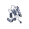
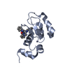
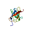
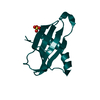
- PDB-3uw4: Crystal structure of cIAP1 BIR3 bound to GDC0152 -
+
Open data
ID or keywords:
Loading...
-
Basic information
Entry
Database: PDB / ID: 3uw4
Title
Crystal structure of cIAP1 BIR3 bound to GDC0152
Components
Baculoviral IAP repeat-containing protein 2, Baculoviral IAP repeat-containing protein 4
GDC0152
Keywords
Apoptosis Inhibitor / BIR domain
Function / homology
Function and homology information
negative regulation of ripoptosome assembly involved in necroptotic process / response to bleomycin / FBXO family protein binding / regulation of RIG-I signaling pathway / positive regulation of protein K48-linked ubiquitination / regulation of apoptosis involved in tissue homeostasis / regulation of non-canonical NF-kappaB signal transduction / positive regulation of protein linear polyubiquitination / TNF receptor superfamily (TNFSF) members mediating non-canonical NF-kB pathway / regulation of BMP signaling pathway ...negative regulation of ripoptosome assembly involved in necroptotic process / response to bleomycin / FBXO family protein binding / regulation of RIG-I signaling pathway / positive regulation of protein K48-linked ubiquitination / regulation of apoptosis involved in tissue homeostasis / regulation of non-canonical NF-kappaB signal transduction / positive regulation of protein linear polyubiquitination / TNF receptor superfamily (TNFSF) members mediating non-canonical NF-kB pathway / regulation of BMP signaling pathway / copper ion homeostasis / nucleotide-binding oligomerization domain containing 1 signaling pathway / regulation of necroptotic process / regulation of nucleotide-binding domain, leucine rich repeat containing receptor signaling pathway / positive regulation of protein K63-linked ubiquitination / CD40 receptor complex / nucleotide-binding oligomerization domain containing 2 signaling pathway / SMAC, XIAP-regulated apoptotic response / negative regulation of necroptotic process / cysteine-type endopeptidase inhibitor activity involved in apoptotic process / Activation of caspases through apoptosome-mediated cleavage / SMAC (DIABLO) binds to IAPs / SMAC(DIABLO)-mediated dissociation of IAP:caspase complexes / Regulation of the apoptosome activity / XY body / quinolinate biosynthetic process / TNFR1-induced proapoptotic signaling / positive regulation of protein monoubiquitination / regulation of reactive oxygen species metabolic process / RIPK1-mediated regulated necrosis / non-canonical NF-kappaB signal transduction / Apoptotic cleavage of cellular proteins / regulation of toll-like receptor signaling pathway / regulation of innate immune response / necroptotic process / regulation of cell differentiation / response to cAMP / cysteine-type endopeptidase inhibitor activity / negative regulation of tumor necrosis factor-mediated signaling pathway / protein K63-linked ubiquitination / positive regulation of type I interferon production / canonical NF-kappaB signal transduction / placenta development / Regulation of PTEN localization / positive regulation of protein ubiquitination / tumor necrosis factor-mediated signaling pathway / TICAM1, RIP1-mediated IKK complex recruitment / protein serine/threonine kinase binding / negative regulation of canonical NF-kappaB signal transduction / IKK complex recruitment mediated by RIP1 / TNFR1-induced NF-kappa-B signaling pathway / ubiquitin binding / Deactivation of the beta-catenin transactivating complex / TNFR2 non-canonical NF-kB pathway / Regulation of TNFR1 signaling / NOD1/2 Signaling Pathway / RING-type E3 ubiquitin transferase / positive regulation of JNK cascade / Regulation of necroptotic cell death / cytoplasmic side of plasma membrane / Wnt signaling pathway / Regulation of PTEN stability and activity / protein polyubiquitination / ubiquitin-protein transferase activity / ubiquitin protein ligase activity / positive regulation of canonical Wnt signaling pathway / regulation of cell population proliferation / transferase activity / protein-folding chaperone binding / regulation of inflammatory response / neuron apoptotic process / regulation of apoptotic process / response to lipopolysaccharide / response to ethanol / proteasome-mediated ubiquitin-dependent protein catabolic process / response to hypoxia / transcription coactivator activity / positive regulation of canonical NF-kappaB signal transduction / cell surface receptor signaling pathway / defense response to bacterium / regulation of cell cycle / Ub-specific processing proteases / apoptotic process / DNA damage response / negative regulation of apoptotic process / protein-containing complex binding / zinc ion binding / nucleoplasm / identical protein binding / nucleus / cytoplasm / cytosol Similarity search - Function
BIRC2/BIRC3, UBA domain / XIAP/BIRC8, UBA domain / : / BIRC2/3-like, UBA domain / Inhibitor Of Apoptosis Protein (2mihbC-IAP-1); Chain A / Inhibitor Of Apoptosis Protein (2mihbC-IAP-1); Chain A / : / BIR repeat. / Caspase recruitment domain / BIR repeat ...BIRC2/BIRC3, UBA domain / XIAP/BIRC8, UBA domain / : / BIRC2/3-like, UBA domain / Inhibitor Of Apoptosis Protein (2mihbC-IAP-1); Chain A / Inhibitor Of Apoptosis Protein (2mihbC-IAP-1); Chain A / : / BIR repeat. / Caspase recruitment domain / BIR repeat / Inhibitor of Apoptosis domain / BIR repeat profile. / Baculoviral inhibition of apoptosis protein repeat / CARD domain / CARD caspase recruitment domain profile. / Caspase recruitment domain / Zinc finger, C3HC4 type (RING finger) / Death-like domain superfamily / Ring finger / Zinc finger RING-type profile. / Zinc finger, RING-type / Orthogonal Bundle / Mainly Alpha Similarity search - Domain/homology
Mass: 18.015 Da / Num. of mol.: 53 / Source method: isolated from a natural source / Formula: H2O
-
Experimental details
-
Experiment
Experiment
Method: X-RAY DIFFRACTION
-
Sample preparation
Crystal
Density Matthews: 1.81 Å3/Da / Density % sol: 32.4 %
Crystal grow
Temperature: 292 K / Method: vapor diffusion, hanging drop / pH: 8.6 Details: Protein Solution (4mg/mL cIAP with 1 mM GDC-0152, 50 mM HEPES pH 7.2, 300 mM NaCl, 0.2 mM TCEP) was mixed with equal volumes of resevoir solution (0.1 M Tris-HCl pH 8.6, 0.5 M Magnesium ...Details: Protein Solution (4mg/mL cIAP with 1 mM GDC-0152, 50 mM HEPES pH 7.2, 300 mM NaCl, 0.2 mM TCEP) was mixed with equal volumes of resevoir solution (0.1 M Tris-HCl pH 8.6, 0.5 M Magnesium Formate)., VAPOR DIFFUSION, HANGING DROP, temperature 292K
-
Data collection
Diffraction
Mean temperature: 100 K
Diffraction source
Source: SYNCHROTRON / Site: ALS / Beamline: 5.0.2 / Wavelength: 1 Å
Resolution: 1.79→31.36 Å / Cor.coef. Fo:Fc: 0.958 / Cor.coef. Fo:Fc free: 0.94 / SU B: 5.83 / SU ML: 0.091 / Cross valid method: THROUGHOUT / ESU R Free: 0.142 / Stereochemistry target values: MAXIMUM LIKELIHOOD / Details: HYDROGENS HAVE BEEN USED IF PRESENT IN THE INPUT
Rfactor
Num. reflection
% reflection
Selection details
Rfree
0.22113
736
9.8 %
RANDOM
Rwork
0.17706
-
-
-
all
0.18134
7661
-
-
obs
0.18134
7503
97.92 %
-
Solvent computation
Ion probe radii: 0.8 Å / Shrinkage radii: 0.8 Å / VDW probe radii: 1.4 Å / Solvent model: MASK
Displacement parameters
Biso mean: 21.068 Å2
Baniso -1
Baniso -2
Baniso -3
1-
-1.63 Å2
0 Å2
0 Å2
2-
-
-0.98 Å2
-0 Å2
3-
-
-
2.61 Å2
Refinement step
Cycle: LAST / Resolution: 1.79→31.36 Å
Protein
Nucleic acid
Ligand
Solvent
Total
Num. atoms
746
0
2
53
801
Refine LS restraints
Refine-ID
Type
Dev ideal
Dev ideal target
Number
X-RAY DIFFRACTION
r_bond_refined_d
0.009
0.02
780
X-RAY DIFFRACTION
r_bond_other_d
0.003
0.02
3
X-RAY DIFFRACTION
r_angle_refined_deg
1.37
1.959
1053
X-RAY DIFFRACTION
r_angle_other_deg
1.023
3
9
X-RAY DIFFRACTION
r_dihedral_angle_1_deg
5.15
5
86
X-RAY DIFFRACTION
r_dihedral_angle_2_deg
31.199
23.077
39
X-RAY DIFFRACTION
r_dihedral_angle_3_deg
13.072
15
112
X-RAY DIFFRACTION
r_dihedral_angle_4_deg
14.882
15
5
X-RAY DIFFRACTION
r_chiral_restr
0.089
0.2
99
X-RAY DIFFRACTION
r_gen_planes_refined
0.008
0.021
618
LS refinement shell
Resolution: 1.79→1.827 Å / Total num. of bins used: 25
Rfactor
Num. reflection
% reflection
Rfree
0.296
40
-
Rwork
0.209
325
-
obs
-
-
96.82 %
Refinement TLS params.
Method: refined / Origin x: -6.7745 Å / Origin y: 2.3651 Å / Origin z: -3.9479 Å
11
12
13
21
22
23
31
32
33
T
0.0282 Å2
-0.0062 Å2
0.0095 Å2
-
0.0134 Å2
-0.0051 Å2
-
-
0.009 Å2
L
2.2985 °2
-0.8087 °2
0.6958 °2
-
1.496 °2
-0.6166 °2
-
-
1.2721 °2
S
0.0037 Å °
0.0546 Å °
0.0889 Å °
0.0238 Å °
-0.0242 Å °
-0.0263 Å °
-0.0054 Å °
0.0322 Å °
0.0205 Å °
+
About Yorodumi
-
News
-
Feb 9, 2022. New format data for meta-information of EMDB entries
New format data for meta-information of EMDB entries
Version 3 of the EMDB header file is now the official format.
The previous official version 1.9 will be removed from the archive.
In the structure databanks used in Yorodumi, some data are registered as the other names, "COVID-19 virus" and "2019-nCoV". Here are the details of the virus and the list of structure data.
Jan 31, 2019. EMDB accession codes are about to change! (news from PDBe EMDB page)
EMDB accession codes are about to change! (news from PDBe EMDB page)
The allocation of 4 digits for EMDB accession codes will soon come to an end. Whilst these codes will remain in use, new EMDB accession codes will include an additional digit and will expand incrementally as the available range of codes is exhausted. The current 4-digit format prefixed with “EMD-” (i.e. EMD-XXXX) will advance to a 5-digit format (i.e. EMD-XXXXX), and so on. It is currently estimated that the 4-digit codes will be depleted around Spring 2019, at which point the 5-digit format will come into force.
The EM Navigator/Yorodumi systems omit the EMD- prefix.
Related info.:Q: What is EMD? / ID/Accession-code notation in Yorodumi/EM Navigator
Yorodumi is a browser for structure data from EMDB, PDB, SASBDB, etc.
This page is also the successor to EM Navigator detail page, and also detail information page/front-end page for Omokage search.
The word "yorodu" (or yorozu) is an old Japanese word meaning "ten thousand". "mi" (miru) is to see.
Related info.:EMDB / PDB / SASBDB / Comparison of 3 databanks / Yorodumi Search / Aug 31, 2016. New EM Navigator & Yorodumi / Yorodumi Papers / Jmol/JSmol / Function and homology information / Changes in new EM Navigator and Yorodumi
 Movie
Movie Controller


 Open data
 Basic information
 Components
 Keywords
 Function and homology information
 Homo sapiens (human)
 X-RAY DIFFRACTION /
  Authors
 Citation
 Structure visualization
 Downloads & links
 Other downloads
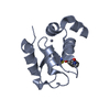


 PDBj
PDBj


 Assembly


 Type: Peptide-like / Class: Inhibitor / Mass: 498.640 Da / Num. of mol.: 1 / Source method: obtained synthetically / Details: Synthetic inhibitor / References: GDC0152
 Mass: 65.409 Da / Num. of mol.: 2 / Source method: obtained synthetically / Formula: Zn
 Mass: 18.015 Da / Num. of mol.: 53 / Source method: isolated from a natural source / Formula: H2O
 Sample preparation
 / Beamline: 5.0.2 / Wavelength: 1 Å
 Processing