[English] 日本語
Yorodumi
- PDB-3ucy: Structure of Mg2+ bound N-terminal domain of calmodulin in the pr... -

+
Open data


ID or keywords:

Loading...

-
Basic information

Entry
Database: PDB / ID: 3ucy
TitleStructure of Mg2+ bound N-terminal domain of calmodulin in the presence of Zn2+
ComponentsCalmodulin
KeywordsCALCIUM-BINDING PROTEIN / EF-HAND / METAL BINDING / CALCIUM REGULATION
Function / homology
Function and homology information


: / establishment of protein localization to mitochondrial membrane / type 3 metabotropic glutamate receptor binding / CaM pathway / response to corticosterone / Cam-PDE 1 activation / Sodium/Calcium exchangers / Calmodulin induced events / Reduction of cytosolic Ca++ levels / Activation of Ca-permeable Kainate Receptor ...: / establishment of protein localization to mitochondrial membrane / type 3 metabotropic glutamate receptor binding / CaM pathway / response to corticosterone / Cam-PDE 1 activation / Sodium/Calcium exchangers / Calmodulin induced events / Reduction of cytosolic Ca++ levels / Activation of Ca-permeable Kainate Receptor / regulation of synaptic vesicle endocytosis / CREB1 phosphorylation through the activation of CaMKII/CaMKK/CaMKIV cascasde / Loss of phosphorylation of MECP2 at T308 / PKA activation / CREB1 phosphorylation through the activation of Adenylate Cyclase / negative regulation of high voltage-gated calcium channel activity / CaMK IV-mediated phosphorylation of CREB / Glycogen breakdown (glycogenolysis) / regulation of synaptic vesicle exocytosis / negative regulation of calcium ion export across plasma membrane / organelle localization by membrane tethering / Activation of RAC1 downstream of NMDARs / regulation of cardiac muscle cell action potential / mitochondrion-endoplasmic reticulum membrane tethering / CLEC7A (Dectin-1) induces NFAT activation / autophagosome membrane docking / Negative regulation of NMDA receptor-mediated neuronal transmission / positive regulation of ryanodine-sensitive calcium-release channel activity / nitric-oxide synthase binding / Unblocking of NMDA receptors, glutamate binding and activation / regulation of cell communication by electrical coupling involved in cardiac conduction / negative regulation of peptidyl-threonine phosphorylation / Synthesis of IP3 and IP4 in the cytosol / Phase 0 - rapid depolarisation / protein phosphatase activator activity / RHO GTPases activate PAKs / positive regulation of cyclic-nucleotide phosphodiesterase activity / Long-term potentiation / positive regulation of phosphoprotein phosphatase activity / Ion transport by P-type ATPases / Uptake and function of anthrax toxins / Regulation of MECP2 expression and activity / Calcineurin activates NFAT / adenylate cyclase binding / catalytic complex / detection of calcium ion / DARPP-32 events / Smooth Muscle Contraction / negative regulation of ryanodine-sensitive calcium-release channel activity / RHO GTPases activate IQGAPs / cellular response to interferon-beta / regulation of cardiac muscle contraction / calcium channel inhibitor activity / regulation of cardiac muscle contraction by regulation of the release of sequestered calcium ion / positive regulation of DNA binding / Protein methylation / enzyme regulator activity / voltage-gated potassium channel complex / Activation of AMPK downstream of NMDARs / phosphatidylinositol 3-kinase binding / eNOS activation / regulation of release of sequestered calcium ion into cytosol by sarcoplasmic reticulum / response to amphetamine / regulation of calcium-mediated signaling / positive regulation of protein dephosphorylation / titin binding / regulation of ryanodine-sensitive calcium-release channel activity / Tetrahydrobiopterin (BH4) synthesis, recycling, salvage and regulation / Ion homeostasis / positive regulation of protein autophosphorylation / sperm midpiece / calcium channel complex / activation of adenylate cyclase activity / substantia nigra development / adenylate cyclase activator activity / Ras activation upon Ca2+ influx through NMDA receptor / protein serine/threonine kinase activator activity / regulation of heart rate / nitric-oxide synthase regulator activity / sarcomere / FCERI mediated Ca+2 mobilization / FCGR3A-mediated IL10 synthesis / VEGFR2 mediated vascular permeability / positive regulation of peptidyl-threonine phosphorylation / Antigen activates B Cell Receptor (BCR) leading to generation of second messengers / regulation of cytokinesis / VEGFR2 mediated cell proliferation / positive regulation of nitric-oxide synthase activity / Translocation of SLC2A4 (GLUT4) to the plasma membrane / spindle microtubule / mitochondrial membrane / RAF activation / positive regulation of receptor signaling pathway via JAK-STAT / positive regulation of protein serine/threonine kinase activity / Transcriptional activation of mitochondrial biogenesis / synaptic vesicle membrane / cellular response to type II interferon / Stimuli-sensing channels / spindle pole / response to calcium ion
Similarity search - Function
EF-hand domain pair / EF-hand, calcium binding motif / EF-Hand 1, calcium-binding site / EF-hand calcium-binding domain. / EF-hand calcium-binding domain profile. / EF-hand domain / EF-hand domain pair
Similarity search - Domain/homology
CACODYLATE ION / Calmodulin-1 / Calmodulin-3
Similarity search - Component
Biological speciesHomo sapiens (human)
MethodX-RAY DIFFRACTION / SYNCHROTRON / SAD / Resolution: 1.8 Å
AuthorsSenguen, F.T. / Grabarek, Z.
Citation
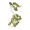
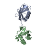
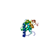
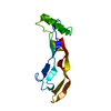
Journal: Biochemistry / Year: 2012
Title: X-ray Structures of Magnesium and Manganese Complexes with the N-Terminal Domain of Calmodulin: Insights into the Mechanism and Specificity of Metal Ion Binding to an EF-Hand.
Authors: Senguen, F.T. / Grabarek, Z.
History
DepositionOct 27, 2011Deposition site: RCSB / Processing site: RCSB
Revision 1.0Aug 8, 2012Provider: repository / Type: Initial release
Revision 1.1Oct 3, 2012Group: Database references
Revision 1.2Feb 28, 2024Group: Data collection / Database references / Derived calculations
Category: chem_comp_atom / chem_comp_bond ...chem_comp_atom / chem_comp_bond / database_2 / pdbx_struct_conn_angle / struct_conn / struct_site
Item: _database_2.pdbx_DOI / _database_2.pdbx_database_accession ..._database_2.pdbx_DOI / _database_2.pdbx_database_accession / _pdbx_struct_conn_angle.ptnr1_auth_comp_id / _pdbx_struct_conn_angle.ptnr1_auth_seq_id / _pdbx_struct_conn_angle.ptnr1_label_asym_id / _pdbx_struct_conn_angle.ptnr1_label_atom_id / _pdbx_struct_conn_angle.ptnr1_label_comp_id / _pdbx_struct_conn_angle.ptnr1_label_seq_id / _pdbx_struct_conn_angle.ptnr2_auth_comp_id / _pdbx_struct_conn_angle.ptnr2_auth_seq_id / _pdbx_struct_conn_angle.ptnr2_label_asym_id / _pdbx_struct_conn_angle.ptnr2_label_atom_id / _pdbx_struct_conn_angle.ptnr2_label_comp_id / _pdbx_struct_conn_angle.ptnr3_auth_comp_id / _pdbx_struct_conn_angle.ptnr3_auth_seq_id / _pdbx_struct_conn_angle.ptnr3_label_asym_id / _pdbx_struct_conn_angle.ptnr3_label_atom_id / _pdbx_struct_conn_angle.ptnr3_label_comp_id / _pdbx_struct_conn_angle.ptnr3_label_seq_id / _pdbx_struct_conn_angle.value / _struct_conn.pdbx_dist_value / _struct_conn.ptnr1_auth_comp_id / _struct_conn.ptnr1_auth_seq_id / _struct_conn.ptnr1_label_asym_id / _struct_conn.ptnr1_label_atom_id / _struct_conn.ptnr1_label_comp_id / _struct_conn.ptnr1_label_seq_id / _struct_conn.ptnr2_auth_comp_id / _struct_conn.ptnr2_auth_seq_id / _struct_conn.ptnr2_label_asym_id / _struct_conn.ptnr2_label_atom_id / _struct_conn.ptnr2_label_comp_id / _struct_site.pdbx_auth_asym_id / _struct_site.pdbx_auth_comp_id / _struct_site.pdbx_auth_seq_id

-
Structure visualization

Structure viewerMolecule:
MolmilJmol/JSmol

Downloads & links

-
Assembly

Deposited unit
A: Calmodulin
hetero molecules


Theoretical massNumber of molelcules
Total (without water)9,1156
Polymers8,8001
Non-polymers3155
Water1,22568
1
A: Calmodulin
hetero molecules

A: Calmodulin
hetero molecules


Theoretical massNumber of molelcules
Total (without water)18,23012
Polymers17,5992
Non-polymers63010
Water362
TypeNameSymmetry operationNumber
identity operation1_555x,y,z1
crystal symmetry operation7_556y,x,-z+11
Buried area1950 Å2
ΔGint-159 kcal/mol
Surface area8850 Å2
MethodPISA
Unit cell
Length a, b, c (Å)35.298, 35.298, 142.937
Angle α, β, γ (deg.)90.00, 90.00, 90.00
Int Tables number96
Space group name H-MP43212

-
Components

-
Protein , 1 types, 1 molecules A

#1: Protein Calmodulin / / CaM


Mass: 8799.724 Da / Num. of mol.: 1 / Fragment: N-terminal domain residues 2-80
Source method: isolated from a genetically manipulated source
Source: (gene. exp.) Homo sapiens (human)
Gene: CALM, CALM1, CALM2, CALM3, CALML2, CAM, CAM1, CAM2, CAM3, CAMB, CAMC, CAMIII
Plasmid: PAED4 / Production host: Escherichia coli (E. coli) / Strain (production host): BL21 / References: UniProt: P62158, UniProt: P0DP23*PLUS

-
Non-polymers , 5 types, 73 molecules

#2: Chemical ChemComp-NA / SODIUM ION


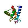
Mass: 22.990 Da / Num. of mol.: 1 / Source method: obtained synthetically / Formula: Na
#3: Chemical ChemComp-MG / MAGNESIUM ION


Mass: 24.305 Da / Num. of mol.: 1 / Source method: obtained synthetically / Formula: Mg
#4: Chemical ChemComp-ZN / ZINC ION


Mass: 65.409 Da / Num. of mol.: 2 / Source method: obtained synthetically / Formula: Zn
#5: Chemical ChemComp-CAC / CACODYLATE ION / dimethylarsinate / Cacodylic acid


Mass: 136.989 Da / Num. of mol.: 1 / Source method: obtained synthetically / Formula: C2H6AsO2
#6: Water ChemComp-HOH / water / Water


Mass: 18.015 Da / Num. of mol.: 68 / Source method: isolated from a natural source / Formula: H2O

-
Experimental details

-
Experiment

ExperimentMethod: X-RAY DIFFRACTION / Number of used crystals: 1

-
Sample preparation

CrystalDensity Matthews: 2.53 Å3/Da / Density % sol: 51.38 %
Crystal growTemperature: 298 K / Method: vapor diffusion, hanging drop / pH: 6.5
Details: 100MM TRIS CACODYLATE, 25MM ZNCL2, 16% PEG8000, 100MM MGCL2, 20% ETHYLENE GLYCOL, pH 6.5, VAPOR DIFFUSION, HANGING DROP, temperature 298K

-
Data collection

DiffractionMean temperature: 100 K
Diffraction sourceSource: SYNCHROTRON / Site: NSLS / Beamline: X29A / Wavelength: 1.075 Å
DetectorType: ADSC QUANTUM 315r / Detector: CCD / Date: Jul 20, 2011 / Details: NINE QUADRANT
RadiationMonochromator: SI(111) / Protocol: SINGLE WAVELENGTH / Monochromatic (M) / Laue (L): M / Scattering type: x-ray
Radiation wavelengthWavelength: 1.075 Å / Relative weight: 1
ReflectionRedundancy: 27.4 % / Av σ(I) over netI: 18.29 / Number: 251118 / Rmerge(I) obs: 0.157 / Χ2: 1.33 / D res high: 1.8 Å / D res low: 50 Å / Num. obs: 9179 / % possible obs: 100
Diffraction reflection shell
Highest resolution (Å)Lowest resolution (Å)% possible obs (%)IDRmerge(I) obsChi squaredRedundancy
3.885099.910.1451.24828.1
3.083.8810010.1421.08632.7
2.693.0899.810.151.18626.4
2.442.6910010.1751.4125.9
2.272.4410010.2061.56426.4
2.132.2710010.2491.57726.7
2.032.1310010.3071.50226.7
1.942.0310010.371.40327
1.861.9410010.4631.24826.7
1.81.8610010.5921.11926.4
ReflectionResolution: 1.8→50 Å / Num. obs: 9179 / % possible obs: 100 % / Observed criterion σ(I): 2 / Redundancy: 27.4 % / Biso Wilson estimate: 31.869 Å2 / Rmerge(I) obs: 0.157 / Χ2: 1.325 / Net I/σ(I): 12.9
Reflection shell
Resolution (Å)Redundancy (%)Rmerge(I) obsNum. unique allΧ2Diffraction-ID% possible all
1.8-1.8626.40.5928911.1191100
1.86-1.9426.70.4638731.2481100
1.94-2.03270.378761.4031100
2.03-2.1326.70.3078951.5021100
2.13-2.2726.70.2499051.5771100
2.27-2.4426.40.2068851.5641100
2.44-2.6925.90.1759151.411100
2.69-3.0826.40.159251.186199.8
3.08-3.8832.70.1429581.0861100
3.88-5028.10.14510561.248199.9

-
Processing

Software
NameVersionClassificationNB
MOSFLMdata reduction
SCALEPACKdata scaling
REFMAC5.6.0117refinement
PDB_EXTRACT3.1data extraction
DENZOdata reduction
RefinementMethod to determine structure: SAD / Resolution: 1.8→35.73 Å / Cor.coef. Fo:Fc: 0.963 / Cor.coef. Fo:Fc free: 0.965 / Occupancy max: 1 / Occupancy min: 0.1 / SU B: 3.259 / SU ML: 0.095 / Cross valid method: THROUGHOUT / σ(F): 0 / ESU R Free: 0.117 / Stereochemistry target values: MAXIMUM LIKELIHOOD
Details: HYDROGENS HAVE BEEN USED IF PRESENT IN THE INPUT U VALUES: REFINED INDIVIDUALLY
RfactorNum. reflection% reflectionSelection details
Rfree0.2279 433 4.8 %RANDOM
Rwork0.201 ---
obs0.2023 9098 99.75 %-
Solvent computationIon probe radii: 0.8 Å / Shrinkage radii: 0.8 Å / VDW probe radii: 1.2 Å / Solvent model: MASK
Displacement parametersBiso max: 79.96 Å2 / Biso mean: 38.4893 Å2 / Biso min: 23.14 Å2
Baniso -1Baniso -2Baniso -3
1--0.74 Å20 Å20 Å2
2---0.74 Å2-0 Å2
3---1.49 Å2
Refinement stepCycle: LAST / Resolution: 1.8→35.73 Å
ProteinNucleic acidLigandSolventTotal
Num. atoms575 0 9 68 652
Refine LS restraints
Refine-IDTypeDev idealDev ideal targetNumber
X-RAY DIFFRACTIONr_bond_refined_d0.0080.02620
X-RAY DIFFRACTIONr_angle_refined_deg1.3021.985838
X-RAY DIFFRACTIONr_dihedral_angle_1_deg4.417583
X-RAY DIFFRACTIONr_dihedral_angle_2_deg30.86626.56332
X-RAY DIFFRACTIONr_dihedral_angle_3_deg15.86115125
X-RAY DIFFRACTIONr_dihedral_angle_4_deg12.745153
X-RAY DIFFRACTIONr_chiral_restr0.0960.293
X-RAY DIFFRACTIONr_gen_planes_refined0.0050.021466
LS refinement shellResolution: 1.8→1.844 Å / Total num. of bins used: 20
RfactorNum. reflection% reflection
Rfree0.364 29 -
Rwork0.243 517 -
all-546 -
obs--99.27 %

+
About Yorodumi

-
News

-
Feb 9, 2022. New format data for meta-information of EMDB entries

New format data for meta-information of EMDB entries

  • Version 3 of the EMDB header file is now the official format.
  • The previous official version 1.9 will be removed from the archive.

Related info.:EMDB header

External links:wwPDB to switch to version 3 of the EMDB data model

-
Aug 12, 2020. Covid-19 info

Covid-19 info

URL: https://pdbj.org/emnavi/covid19.php

New page: Covid-19 featured information page in EM Navigator.

Related info.:Covid-19 info / Mar 5, 2020. Novel coronavirus structure data

+
Mar 5, 2020. Novel coronavirus structure data

Novel coronavirus structure data

Related info.:Yorodumi Speices / Aug 12, 2020. Covid-19 info

External links:COVID-19 featured content - PDBj / Molecule of the Month (242):Coronavirus Proteases

+
Jan 31, 2019. EMDB accession codes are about to change! (news from PDBe EMDB page)

EMDB accession codes are about to change! (news from PDBe EMDB page)

  • The allocation of 4 digits for EMDB accession codes will soon come to an end. Whilst these codes will remain in use, new EMDB accession codes will include an additional digit and will expand incrementally as the available range of codes is exhausted. The current 4-digit format prefixed with “EMD-” (i.e. EMD-XXXX) will advance to a 5-digit format (i.e. EMD-XXXXX), and so on. It is currently estimated that the 4-digit codes will be depleted around Spring 2019, at which point the 5-digit format will come into force.
  • The EM Navigator/Yorodumi systems omit the EMD- prefix.

Related info.:Q: What is EMD? / ID/Accession-code notation in Yorodumi/EM Navigator

External links:EMDB Accession Codes are Changing Soon! / Contact to PDBj

+
Jul 12, 2017. Major update of PDB

Major update of PDB

  • wwPDB released updated PDB data conforming to the new PDBx/mmCIF dictionary.
  • This is a major update changing the version number from 4 to 5, and with Remediation, in which all the entries are updated.
  • In this update, many items about electron microscopy experimental information are reorganized (e.g. em_software).
  • Now, EM Navigator and Yorodumi are based on the updated data.

External links:wwPDB Remediation / Enriched Model Files Conforming to OneDep Data Standards Now Available in the PDB FTP Archive

-
Yorodumi

Thousand views of thousand structures

  • Yorodumi is a browser for structure data from EMDB, PDB, SASBDB, etc.
  • This page is also the successor to EM Navigator detail page, and also detail information page/front-end page for Omokage search.
  • The word "yorodu" (or yorozu) is an old Japanese word meaning "ten thousand". "mi" (miru) is to see.

Related info.:EMDB / PDB / SASBDB / Comparison of 3 databanks / Yorodumi Search / Aug 31, 2016. New EM Navigator & Yorodumi / Yorodumi Papers / Jmol/JSmol / Function and homology information / Changes in new EM Navigator and Yorodumi

Read more