 Movie
Movie Controller

[English] 日本語
 Yorodumi
- PDB-3u6r: Three dimensional structure of broadly neutralizing anti - Hepati... -
+ Open data

- Basic information
| Entry | Database: PDB / ID: 3u6r | ||||||
|---|---|---|---|---|---|---|---|
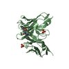
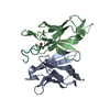
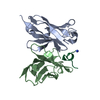
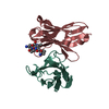
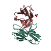
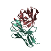
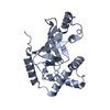
| Title | Three dimensional structure of broadly neutralizing anti - Hepatitis C virus (HCV) glycoprotein E2 single chain FV fragment 1:7 | ||||||
 Components Components |
| ||||||
 Keywords Keywords | IMMUNE SYSTEM / Ig-like domain / neutralizing single chain FV | ||||||
| Function / homology | Immunoglobulins / Immunoglobulin-like / Sandwich / Mainly Beta Function and homology information Function and homology information | ||||||
| Biological species |  Homo sapiens (human) Homo sapiens (human) | ||||||
| Method |  X-RAY DIFFRACTION / SYNCHROTRON / MOLECULAR REPLACEMENT / Resolution: 2.67 Å X-RAY DIFFRACTION / SYNCHROTRON / MOLECULAR REPLACEMENT / Resolution: 2.67 Å | ||||||
 Authors Authors | Gilmartin, A.A. / Lamp, B. / Ruemenapf, T. / Persson, A.A. / Rey, F.A. / Krey, T. | ||||||
 Citation Citation | Journal: Protein Eng.Des.Sel. / Year: 2012 Title: High-level secretion of recombinant monomeric murine and human single-chain Fv antibodies from Drosophila S2 cells. Authors: Gilmartin, A.A. / Lamp, B. / Rumenapf, T. / Persson, M.A. / Rey, F.A. / Krey, T. | ||||||
| History |
|
- Structure visualization
| Structure viewer | Molecule: MolmilJmol/JSmol |
|---|
- Downloads & links
-Download
| PDBx/mmCIF format | 3u6r.cif.gz | 103.2 KB | Display | PDBx/mmCIF format |
|---|---|---|---|---|
| PDB format | pdb3u6r.ent.gz | 78.7 KB | Display | PDB format |
| PDBx/mmJSON format | 3u6r.json.gz | Tree view | PDBx/mmJSON format | |
| Others |  Other downloads Other downloads |
-Validation report
| Arichive directory | https://data.pdbj.org/pub/pdb/validation_reports/u6/3u6rftp://data.pdbj.org/pub/pdb/validation_reports/u6/3u6r | HTTPS FTP |
|---|
-Related structure data
| Related structure data |  3fkuS S: Starting model for refinement |
|---|---|
| Similar structure data |


-Links
 PDBj
PDBj

- Assembly
| Deposited unit | 
| ||||||||
|---|---|---|---|---|---|---|---|---|---|
| 1 | 
| ||||||||
| 2 | 
| ||||||||
| Unit cell |
|
-Components
| #1: Antibody | Mass: 15723.355 Da / Num. of mol.: 2 Source method: isolated from a genetically manipulated source Source: (gene. exp.) Homo sapiens (human) / Plasmid: pMT-scFv-Strep / Production host:  #2: Antibody | Mass: 15225.559 Da / Num. of mol.: 2 Source method: isolated from a genetically manipulated source Source: (gene. exp.) Homo sapiens (human) / Plasmid: pMT-scFv-Strep / Production host: #3: Chemical | ChemComp-SO4 / |   Mass: 96.063 Da / Num. of mol.: 1 / Source method: obtained synthetically / Formula: SO4 Mass: 96.063 Da / Num. of mol.: 1 / Source method: obtained synthetically / Formula: SO4#4: Water | ChemComp-HOH / |  Mass: 18.015 Da / Num. of mol.: 58 / Source method: isolated from a natural source / Formula: H2O Mass: 18.015 Da / Num. of mol.: 58 / Source method: isolated from a natural source / Formula: H2OHas protein modification | Y | |
|---|
-Experimental details
-Experiment
| Experiment | Method: X-RAY DIFFRACTION / Number of used crystals: 1 |
|---|
- Sample preparation
| Crystal | Density Matthews: 2.38 Å3/Da / Density % sol: 48.29 % |
|---|---|
| Crystal grow | Temperature: 293 K / Method: vapor diffusion, sitting drop / pH: 8.5 Details: 1.54M ammonium sulfate, 0.2M lithium sulfate, 0.1M Tris/HCl, pH 8.5, VAPOR DIFFUSION, SITTING DROP, temperature 293K |
-Data collection
| Diffraction | Mean temperature: 110 K |
|---|---|
| Diffraction source | Source: SYNCHROTRON / Site: SOLEIL  / Beamline: PROXIMA 1 / Wavelength: 0.98011 Å / Beamline: PROXIMA 1 / Wavelength: 0.98011 Å |
| Detector | Type: ADSC QUANTUM 315r / Detector: CCD / Date: Jun 8, 2011 |
| Radiation | Monochromator: Channel cut Si(111) / Protocol: SINGLE WAVELENGTH / Monochromatic (M) / Laue (L): M / Scattering type: x-ray |
| Radiation wavelength | Wavelength: 0.98011 Å / Relative weight: 1 |
| Reflection | Resolution: 2.67→37.36 Å / Num. all: 17118 / Num. obs: 15834 / % possible obs: 92.5 % / Observed criterion σ(I): -3 / Redundancy: 3.5 % / Biso Wilson estimate: 58.77 Å2 |
| Reflection shell | Resolution: 2.67→2.81 Å / Redundancy: 2.4 % / Rmerge(I) obs: 0.459 / Mean I/σ(I) obs: 1.9 / Num. unique all: 1316 / % possible all: 54.1 |
- Processing
| Software |
| ||||||||||||||||||||||||||||||||||||||||||||||||||||||||||||||||||
|---|---|---|---|---|---|---|---|---|---|---|---|---|---|---|---|---|---|---|---|---|---|---|---|---|---|---|---|---|---|---|---|---|---|---|---|---|---|---|---|---|---|---|---|---|---|---|---|---|---|---|---|---|---|---|---|---|---|---|---|---|---|---|---|---|---|---|---|
| Refinement | Method to determine structure: MOLECULAR REPLACEMENT Starting model: PDB ENTRY 3FKU Resolution: 2.67→24.57 Å / Cor.coef. Fo:Fc: 0.9265 / Cor.coef. Fo:Fc free: 0.8905 / SU R Cruickshank DPI: 0.943 / Cross valid method: THROUGHOUT / σ(F): 0 / σ(I): 2 / Stereochemistry target values: Maximum Likelihood
| ||||||||||||||||||||||||||||||||||||||||||||||||||||||||||||||||||
| Displacement parameters | Biso mean: 50.58 Å2
| ||||||||||||||||||||||||||||||||||||||||||||||||||||||||||||||||||
| Refine analyze | Luzzati coordinate error obs: 0.315 Å | ||||||||||||||||||||||||||||||||||||||||||||||||||||||||||||||||||
| Refinement step | Cycle: LAST / Resolution: 2.67→24.57 Å
| ||||||||||||||||||||||||||||||||||||||||||||||||||||||||||||||||||
| Refine LS restraints |
| ||||||||||||||||||||||||||||||||||||||||||||||||||||||||||||||||||
| LS refinement shell | Resolution: 2.67→2.85 Å / Total num. of bins used: 8
|
