 Movie
Movie Controller


+ Open data

- Basic information
| Entry | Database: PDB / ID: 1g24 | ||||||
|---|---|---|---|---|---|---|---|
| Title | THE CRYSTAL STRUCTURE OF EXOENZYME C3 FROM CLOSTRIDIUM BOTULINUM | ||||||
 Components Components | EXOENZYME C3 | ||||||
 Keywords Keywords | TRANSFERASE / C3 / ADP-ribosyltransferase | ||||||
| Function / homology |  Function and homology information Function and homology informationNAD+-protein mono-ADP-ribosyltransferase activity / Transferases; Glycosyltransferases; Pentosyltransferases / nucleotidyltransferase activity / extracellular region Similarity search - Function | ||||||
| Biological species |   Clostridium botulinum (bacteria) Clostridium botulinum (bacteria) | ||||||
| Method |  X-RAY DIFFRACTION / SYNCHROTRON / MOLECULAR REPLACEMENT / Resolution: 1.7 Å X-RAY DIFFRACTION / SYNCHROTRON / MOLECULAR REPLACEMENT / Resolution: 1.7 Å | ||||||
 Authors Authors | Han, S. / Arvai, A.S. / Clancy, S.B. / Tainer, J.A. | ||||||
 Citation Citation | Journal: J.Mol.Biol. / Year: 2001 Title: Crystal structure and novel recognition motif of rho ADP-ribosylating C3 exoenzyme from Clostridium botulinum: structural insights for recognition specificity and catalysis. Authors: Han, S. / Arvai, A.S. / Clancy, S.B. / Tainer, J.A. | ||||||
| History |
|
- Structure visualization
| Structure viewer | Molecule: MolmilJmol/JSmol |
|---|
- Downloads & links
-Download
| PDBx/mmCIF format | 1g24.cif.gz | 191.1 KB | Display | PDBx/mmCIF format |
|---|---|---|---|---|
| PDB format | pdb1g24.ent.gz | 149.8 KB | Display | PDB format |
| PDBx/mmJSON format | 1g24.json.gz | Tree view | PDBx/mmJSON format | |
| Others |  Other downloads Other downloads |
-Validation report
| Arichive directory | https://data.pdbj.org/pub/pdb/validation_reports/g2/1g24ftp://data.pdbj.org/pub/pdb/validation_reports/g2/1g24 | HTTPS FTP |
|---|
-Related structure data
| Related structure data | 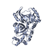 1qs1S S: Starting model for refinement |
|---|---|
| Similar structure data |


-Links
 PDBj
PDBj- Assembly
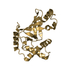
| Deposited unit | 
| ||||||||||
|---|---|---|---|---|---|---|---|---|---|---|---|
| 1 | 
| ||||||||||
| 2 | 
| ||||||||||
| 3 | 
| ||||||||||
| 4 | 
| ||||||||||
| Unit cell |
|
-Components
| #1: Protein | Mass: 23591.969 Da / Num. of mol.: 4 Source method: isolated from a genetically manipulated source Source: (gene. exp.) Clostridium botulinum (bacteria) / Production host: References: UniProt: P15879, Transferases; Glycosyltransferases; Pentosyltransferases #2: Water | ChemComp-HOH / |  Mass: 18.015 Da / Num. of mol.: 869 / Source method: isolated from a natural source / Formula: H2O Mass: 18.015 Da / Num. of mol.: 869 / Source method: isolated from a natural source / Formula: H2O |
|---|
-Experimental details
-Experiment
| Experiment | Method: X-RAY DIFFRACTION |
|---|
- Sample preparation
| Crystal | Density Matthews: 3.22 Å3/Da / Density % sol: 61.8 % | |||||||||||||||||||||||||
|---|---|---|---|---|---|---|---|---|---|---|---|---|---|---|---|---|---|---|---|---|---|---|---|---|---|---|
| Crystal grow | Temperature: 277 K / Method: vapor diffusion, hanging drop / pH: 5 Details: PEG 5000, monomethylether, ethyleneglycol, imidazole, malate, sodium orthovanadate, pH 5.0, VAPOR DIFFUSION, HANGING DROP, temperature 277K | |||||||||||||||||||||||||
| Crystal grow | *PLUS Temperature: 4 ℃ | |||||||||||||||||||||||||
| Components of the solutions | *PLUS
|
-Data collection
| Diffraction | Mean temperature: 100 K |
|---|---|
| Diffraction source | Source: SYNCHROTRON / Site: SSRL  / Beamline: BL9-1 / Wavelength: 1 Å / Beamline: BL9-1 / Wavelength: 1 Å |
| Detector | Type: RIGAKU AFC-5 / Detector: CCD / Date: Apr 20, 1999 |
| Radiation | Protocol: SINGLE WAVELENGTH / Monochromatic (M) / Laue (L): M / Scattering type: x-ray |
| Radiation wavelength | Wavelength: 1 Å / Relative weight: 1 |
| Reflection | Resolution: 1.7→30 Å / Num. all: 124350 / Num. obs: 124350 / Observed criterion σ(F): 0 / Observed criterion σ(I): 0 / Redundancy: 7.7 % / Biso Wilson estimate: 22 Å2 / Rsym value: 5.7 / Net I/σ(I): 6 |
| Reflection shell | Resolution: 1.7→1.76 Å / Redundancy: 3.5 % / Mean I/σ(I) obs: 4.4 / Num. unique all: 12418 / Rsym value: 16.4 |
| Reflection | *PLUS % possible obs: 94.6 % / Num. measured all: 359859 / Rmerge(I) obs: 0.057 |
| Reflection shell | *PLUS % possible obs: 95 % / Rmerge(I) obs: 0.164 |
- Processing
| Software |
| |||||||||||||||||||||||||
|---|---|---|---|---|---|---|---|---|---|---|---|---|---|---|---|---|---|---|---|---|---|---|---|---|---|---|
| Refinement | Method to determine structure: MOLECULAR REPLACEMENT Starting model: PDB ENTRY 1QS1 Resolution: 1.7→30 Å / Cross valid method: THROUGHOUT / σ(F): 0 / σ(I): 0 / Stereochemistry target values: Engh & Huber
| |||||||||||||||||||||||||
| Refinement step | Cycle: LAST / Resolution: 1.7→30 Å
| |||||||||||||||||||||||||
| Refine LS restraints |
|
