 Movie
Movie Controller

[English] 日本語
 Yorodumi
- PDB-3syo: Crystal structure of the G protein-gated inward rectifier K+ chan... -
+ Open data

- Basic information
| Entry | Database: PDB / ID: 3syo | ||||||
|---|---|---|---|---|---|---|---|
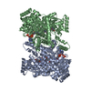
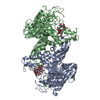
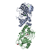
| Title | Crystal structure of the G protein-gated inward rectifier K+ channel GIRK2 (Kir3.2) in complex with sodium | ||||||
 Components Components | G protein-activated inward rectifier potassium channel 2 | ||||||
 Keywords Keywords | METAL TRANSPORT / ion channel / potassium channel / inward rectification / sodium binding / PIP2 binding / G protein binding | ||||||
| Function / homology |  Function and homology information Function and homology informationG-protein activated inward rectifier potassium channel activity / Activation of G protein gated Potassium channels / Inhibition of voltage gated Ca2+ channels via Gbeta/gamma subunits / inward rectifier potassium channel complex / voltage-gated monoatomic ion channel activity involved in regulation of presynaptic membrane potential / inward rectifier potassium channel activity / regulation of monoatomic ion transmembrane transport / neuronal cell body membrane / potassium ion import across plasma membrane / parallel fiber to Purkinje cell synapse ...G-protein activated inward rectifier potassium channel activity / Activation of G protein gated Potassium channels / Inhibition of voltage gated Ca2+ channels via Gbeta/gamma subunits / inward rectifier potassium channel complex / voltage-gated monoatomic ion channel activity involved in regulation of presynaptic membrane potential / inward rectifier potassium channel activity / regulation of monoatomic ion transmembrane transport / neuronal cell body membrane / potassium ion import across plasma membrane / parallel fiber to Purkinje cell synapse / G-protein alpha-subunit binding / potassium channel activity / negative regulation of insulin secretion / presynapse / presynaptic membrane / postsynapse / axon / dendrite / cell surface / membrane / plasma membrane Similarity search - Function | ||||||
| Biological species |  | ||||||
| Method |  X-RAY DIFFRACTION / SYNCHROTRON / MOLECULAR REPLACEMENT / Resolution: 3.54 Å X-RAY DIFFRACTION / SYNCHROTRON / MOLECULAR REPLACEMENT / Resolution: 3.54 Å | ||||||
 Authors Authors | Whorton, M.R. / MacKinnon, R. | ||||||
 Citation Citation | Journal: Cell(Cambridge,Mass.) / Year: 2011 Title: Crystal Structure of the Mammalian GIRK2 K(+) Channel and Gating Regulation by G Proteins, PIP(2), and Sodium. Authors: Whorton, M.R. / Mackinnon, R. | ||||||
| History |
|
- Structure visualization
| Structure viewer | Molecule: MolmilJmol/JSmol |
|---|
- Downloads & links
-Download
| PDBx/mmCIF format | 3syo.cif.gz | 137.5 KB | Display | PDBx/mmCIF format |
|---|---|---|---|---|
| PDB format | pdb3syo.ent.gz | 107.7 KB | Display | PDB format |
| PDBx/mmJSON format | 3syo.json.gz | Tree view | PDBx/mmJSON format | |
| Others |  Other downloads Other downloads |
-Validation report
| Arichive directory | https://data.pdbj.org/pub/pdb/validation_reports/sy/3syoftp://data.pdbj.org/pub/pdb/validation_reports/sy/3syo | HTTPS FTP |
|---|
-Related structure data
| Related structure data |  3syaC  3sycC  3sypC  3syqC  2e4fS C: citing same article ( S: Starting model for refinement |
|---|---|
| Similar structure data |

-Links
 PDBj
PDBj

- Assembly
| Deposited unit | 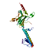
| ||||||||||||||||||
|---|---|---|---|---|---|---|---|---|---|---|---|---|---|---|---|---|---|---|---|
| 1 | 
| ||||||||||||||||||
| Unit cell |
| ||||||||||||||||||
| Components on special symmetry positions |
|
-Components
| #1: Protein | Mass: 39061.957 Da / Num. of mol.: 1 / Fragment: UNP residues 52-380 Source method: isolated from a genetically manipulated source Source: (gene. exp.)  Pichia pastoris (fungus) / Strain (production host): SMD1163 / References: UniProt: P48542 Pichia pastoris (fungus) / Strain (production host): SMD1163 / References: UniProt: P48542 | ||||
|---|---|---|---|---|---|
| #2: Chemical | ChemComp-NA /   Mass: 22.990 Da / Num. of mol.: 1 / Source method: obtained synthetically / Formula: Na Mass: 22.990 Da / Num. of mol.: 1 / Source method: obtained synthetically / Formula: Na | ||||
| #3: Chemical | ChemComp-K /   Mass: 39.098 Da / Num. of mol.: 6 / Source method: obtained synthetically / Formula: K Mass: 39.098 Da / Num. of mol.: 6 / Source method: obtained synthetically / Formula: KHas protein modification | Y | Sequence details | UNP P48542 S260T, I313M, AND M344L ARE NATURAL VARIANTS. | |
-Experimental details
-Experiment
| Experiment | Method: X-RAY DIFFRACTION / Number of used crystals: 1 |
|---|
- Sample preparation
| Crystal | Density Matthews: 4.2 Å3/Da / Density % sol: 70.72 % |
|---|---|
| Crystal grow | Temperature: 293.15 K / Method: vapor diffusion, hanging drop / pH: 6 Details: 50 mM sodium citrate, pH 6.0, 1 M sodium chloride, 30-35% PEG400, VAPOR DIFFUSION, HANGING DROP, temperature 293.15K |
-Data collection
| Diffraction | Mean temperature: 100 K | |||||||||||||||||||||||||||||||||||||||||||||||||||||||||||||||||||||||||||||
|---|---|---|---|---|---|---|---|---|---|---|---|---|---|---|---|---|---|---|---|---|---|---|---|---|---|---|---|---|---|---|---|---|---|---|---|---|---|---|---|---|---|---|---|---|---|---|---|---|---|---|---|---|---|---|---|---|---|---|---|---|---|---|---|---|---|---|---|---|---|---|---|---|---|---|---|---|---|---|
| Diffraction source | Source: SYNCHROTRON / Site: APS  / Beamline: 24-ID-C / Wavelength: 0.9792 Å / Beamline: 24-ID-C / Wavelength: 0.9792 Å | |||||||||||||||||||||||||||||||||||||||||||||||||||||||||||||||||||||||||||||
| Detector | Type: ADSC QUANTUM 315 / Detector: CCD / Date: Dec 13, 2009 | |||||||||||||||||||||||||||||||||||||||||||||||||||||||||||||||||||||||||||||
| Radiation | Monochromator: Rosenbaum-Rock double crystal sagittal focusing Protocol: SINGLE WAVELENGTH / Monochromatic (M) / Laue (L): M / Scattering type: x-ray | |||||||||||||||||||||||||||||||||||||||||||||||||||||||||||||||||||||||||||||
| Radiation wavelength | Wavelength: 0.9792 Å / Relative weight: 1 | |||||||||||||||||||||||||||||||||||||||||||||||||||||||||||||||||||||||||||||
| Reflection | Resolution: 3.54→41.685 Å / Num. obs: 7975 / % possible obs: 92 % / Redundancy: 8.1 % / Rmerge(I) obs: 0.158 / Χ2: 1.605 / Net I/σ(I): 6.4 | |||||||||||||||||||||||||||||||||||||||||||||||||||||||||||||||||||||||||||||
| Reflection shell |
|
- Processing
| Software |
| |||||||||||||||||||||||||||||||||||||||||||||||||||||||||||||||||
|---|---|---|---|---|---|---|---|---|---|---|---|---|---|---|---|---|---|---|---|---|---|---|---|---|---|---|---|---|---|---|---|---|---|---|---|---|---|---|---|---|---|---|---|---|---|---|---|---|---|---|---|---|---|---|---|---|---|---|---|---|---|---|---|---|---|---|
| Refinement | Method to determine structure: MOLECULAR REPLACEMENT Starting model: PDB ENTRY 2E4F Resolution: 3.54→41.68 Å / Cor.coef. Fo:Fc: 0.879 / Cor.coef. Fo:Fc free: 0.905 / Occupancy max: 1 / Occupancy min: 0.13 / SU B: 55.693 / SU ML: 0.404 / Cross valid method: THROUGHOUT / σ(F): 0 / ESU R Free: 0.58 / Stereochemistry target values: MAXIMUM LIKELIHOOD Details: HYDROGENS HAVE BEEN ADDED IN THE RIDING POSITIONS U VALUES : WITH TLS ADDED
| |||||||||||||||||||||||||||||||||||||||||||||||||||||||||||||||||
| Solvent computation | Ion probe radii: 0.8 Å / Shrinkage radii: 0.8 Å / VDW probe radii: 1.4 Å / Solvent model: MASK | |||||||||||||||||||||||||||||||||||||||||||||||||||||||||||||||||
| Displacement parameters | Biso max: 315.82 Å2 / Biso mean: 140.4722 Å2 / Biso min: 68.95 Å2
| |||||||||||||||||||||||||||||||||||||||||||||||||||||||||||||||||
| Refinement step | Cycle: LAST / Resolution: 3.54→41.68 Å
| |||||||||||||||||||||||||||||||||||||||||||||||||||||||||||||||||
| Refine LS restraints |
| |||||||||||||||||||||||||||||||||||||||||||||||||||||||||||||||||
| LS refinement shell | Resolution: 3.544→3.635 Å / Total num. of bins used: 20
| |||||||||||||||||||||||||||||||||||||||||||||||||||||||||||||||||
| Refinement TLS params. | Method: refined / Origin x: -9.6666 Å / Origin y: 30.9521 Å / Origin z: -36.7835 Å
|
