 Movie
Movie Controller

[English] 日本語
 Yorodumi
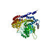
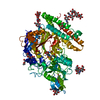
- PDB-2xiq: Crystal structure of human methylmalonyl-CoA mutase in complex wi... -
+ Open data

- Basic information
| Entry | Database: PDB / ID: 2xiq | ||||||
|---|---|---|---|---|---|---|---|
| Title | Crystal structure of human methylmalonyl-CoA mutase in complex with adenosylcobalamin and malonyl-CoA | ||||||
 Components Components | METHYLMALONYL-COA MUTASE, MITOCHONDRIAL | ||||||
 Keywords Keywords | ISOMERASE / ORGANIC ACIDURIA / METABOLIC DISEASE / VITAMIN B12 | ||||||
| Function / homology |  Function and homology information Function and homology informationsuccinyl-CoA biosynthetic process / Defective MMAA causes MMA, cblA type / Defective MUT causes MMAM / : / methylmalonyl-CoA mutase / methylmalonyl-CoA mutase activity / Cobalamin (Cbl) metabolism / Propionyl-CoA catabolism / modified amino acid binding / homocysteine metabolic process ...succinyl-CoA biosynthetic process / Defective MMAA causes MMA, cblA type / Defective MUT causes MMAM / : / methylmalonyl-CoA mutase / methylmalonyl-CoA mutase activity / Cobalamin (Cbl) metabolism / Propionyl-CoA catabolism / modified amino acid binding / homocysteine metabolic process / cobalamin binding / positive regulation of GTPase activity / post-embryonic development / mitochondrial matrix / GTPase activity / protein homodimerization activity / mitochondrion / metal ion binding / identical protein binding / cytoplasm Similarity search - Function | ||||||
| Biological species |  HOMO SAPIENS (human) HOMO SAPIENS (human) | ||||||
| Method |  X-RAY DIFFRACTION / SYNCHROTRON / MOLECULAR REPLACEMENT / Resolution: 1.95 Å X-RAY DIFFRACTION / SYNCHROTRON / MOLECULAR REPLACEMENT / Resolution: 1.95 Å | ||||||
 Authors Authors | Yue, W.W. / Froese, D.S. / Kochan, G. / Chaikuad, A. / Krojer, T. / Muniz, J. / Ugochukwu, E. / Arrowsmith, C. / Weigelt, J. / Edwards, A. ...Yue, W.W. / Froese, D.S. / Kochan, G. / Chaikuad, A. / Krojer, T. / Muniz, J. / Ugochukwu, E. / Arrowsmith, C. / Weigelt, J. / Edwards, A. / Bountra, C. / Oppermann, U. | ||||||
 Citation Citation | Journal: J.Biol.Chem. / Year: 2010 Title: Structures of the Human Gtpase Mmaa and Vitamin B12-Dependent Methylmalonyl-Coa Mutase and Insight Into Their Complex Formation. Authors: Froese, D.S. / Kochan, G. / Muniz, J. / Wu, X. / Gileadi, C. / Ugochukwu, E. / Krysztofinska, E. / Gravel, R.A. / Oppermann, U. / Yue, W.W. | ||||||
| History |
|
- Structure visualization
| Structure viewer | Molecule: MolmilJmol/JSmol |
|---|
- Downloads & links
-Download
| PDBx/mmCIF format | 2xiq.cif.gz | 590.8 KB | Display | PDBx/mmCIF format |
|---|---|---|---|---|
| PDB format | pdb2xiq.ent.gz | 486.2 KB | Display | PDB format |
| PDBx/mmJSON format | 2xiq.json.gz | Tree view | PDBx/mmJSON format | |
| Others |  Other downloads Other downloads |
-Validation report
| Arichive directory | https://data.pdbj.org/pub/pdb/validation_reports/xi/2xiqftp://data.pdbj.org/pub/pdb/validation_reports/xi/2xiq | HTTPS FTP |
|---|
-Related structure data
| Related structure data | 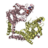 2wwwC 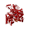 2xijC  3bicSC C: citing same article ( S: Starting model for refinement |
|---|---|
| Similar structure data |


-Links
 PDBj
PDBj- Assembly
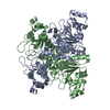
| Deposited unit | 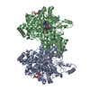
| ||||||||
|---|---|---|---|---|---|---|---|---|---|
| 1 | 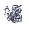
| ||||||||
| 2 | 
| ||||||||
| 3 | 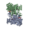
| ||||||||
| Unit cell |
|
-Components
| #1: Protein | Mass: 84835.820 Da / Num. of mol.: 2 / Fragment: RESIDUES 12-750 Source method: isolated from a genetically manipulated source Source: (gene. exp.) HOMO SAPIENS (human) / Production host:  #2: Chemical |   Mass: 1330.356 Da / Num. of mol.: 2 / Source method: obtained synthetically / Formula: C62H89CoN13O14P Mass: 1330.356 Da / Num. of mol.: 2 / Source method: obtained synthetically / Formula: C62H89CoN13O14P#3: Chemical |   Mass: 251.242 Da / Num. of mol.: 2 / Source method: obtained synthetically / Formula: C10H13N5O3 Mass: 251.242 Da / Num. of mol.: 2 / Source method: obtained synthetically / Formula: C10H13N5O3#4: Chemical |   Mass: 853.580 Da / Num. of mol.: 2 / Source method: obtained synthetically / Formula: C24H38N7O19P3S Mass: 853.580 Da / Num. of mol.: 2 / Source method: obtained synthetically / Formula: C24H38N7O19P3S#5: Water | ChemComp-HOH / |  Mass: 18.015 Da / Num. of mol.: 1106 / Source method: isolated from a natural source / Formula: H2O Mass: 18.015 Da / Num. of mol.: 1106 / Source method: isolated from a natural source / Formula: H2O |
|---|
-Experimental details
-Experiment
| Experiment | Method: X-RAY DIFFRACTION / Number of used crystals: 1 |
|---|
- Sample preparation
| Crystal | Density Matthews: 2.75 Å3/Da / Density % sol: 55.3 % / Description: NONE |
|---|---|
| Crystal grow | Details: 20% PEG3350, 0.1 M BIS-TRIS PH 5.5, 0.1 M NH4-SULPHATE |
-Data collection
| Diffraction | Mean temperature: 100 K |
|---|---|
| Diffraction source | Source: SYNCHROTRON / Site: Diamond  / Beamline: I02 / Wavelength: 0.9824 / Beamline: I02 / Wavelength: 0.9824 |
| Detector | Type: ADSC CCD / Detector: CCD / Date: May 9, 2010 |
| Radiation | Monochromator: SI (111) / Protocol: SINGLE WAVELENGTH / Monochromatic (M) / Laue (L): M / Scattering type: x-ray |
| Radiation wavelength | Wavelength: 0.9824 Å / Relative weight: 1 |
| Reflection | Resolution: 1.95→53.91 Å / Num. obs: 129983 / % possible obs: 99.3 % / Observed criterion σ(I): 2 / Redundancy: 4.4 % / Rmerge(I) obs: 0.11 / Net I/σ(I): 9.8 |
| Reflection shell | Resolution: 1.95→2.06 Å / Redundancy: 4.1 % / Rmerge(I) obs: 0.68 / Mean I/σ(I) obs: 2 / % possible all: 97.3 |
- Processing
| Software |
| ||||||||||||||||||||||||||||||||||||||||||||||||||||||||||||||||||||||||||||||||||||||||||||||||||||||||||||||||||||||||||||||||||||||||||||||||||||||||||||||||||||||||||||||||||||||
|---|---|---|---|---|---|---|---|---|---|---|---|---|---|---|---|---|---|---|---|---|---|---|---|---|---|---|---|---|---|---|---|---|---|---|---|---|---|---|---|---|---|---|---|---|---|---|---|---|---|---|---|---|---|---|---|---|---|---|---|---|---|---|---|---|---|---|---|---|---|---|---|---|---|---|---|---|---|---|---|---|---|---|---|---|---|---|---|---|---|---|---|---|---|---|---|---|---|---|---|---|---|---|---|---|---|---|---|---|---|---|---|---|---|---|---|---|---|---|---|---|---|---|---|---|---|---|---|---|---|---|---|---|---|---|---|---|---|---|---|---|---|---|---|---|---|---|---|---|---|---|---|---|---|---|---|---|---|---|---|---|---|---|---|---|---|---|---|---|---|---|---|---|---|---|---|---|---|---|---|---|---|---|---|
| Refinement | Method to determine structure: MOLECULAR REPLACEMENT Starting model: PDB ENTRY 3BIC Resolution: 1.95→107.82 Å / Cor.coef. Fo:Fc: 0.969 / Cor.coef. Fo:Fc free: 0.953 / SU B: 6.371 / SU ML: 0.098 / Cross valid method: THROUGHOUT / σ(F): 2 / ESU R: 0.133 / ESU R Free: 0.128 / Stereochemistry target values: MAXIMUM LIKELIHOOD Details: HYDROGENS HAVE BEEN ADDED IN THE RIDING POSITIONS.ATOM RECORD CONTAINS SUM OF TLS AND RESIDUAL B FACTORS. ANISOU RECORD CONTAINS SUM OF TLS AND RESIDUAL U FACTORS
| ||||||||||||||||||||||||||||||||||||||||||||||||||||||||||||||||||||||||||||||||||||||||||||||||||||||||||||||||||||||||||||||||||||||||||||||||||||||||||||||||||||||||||||||||||||||
| Solvent computation | Ion probe radii: 0.8 Å / Shrinkage radii: 0.8 Å / VDW probe radii: 1.2 Å / Solvent model: MASK | ||||||||||||||||||||||||||||||||||||||||||||||||||||||||||||||||||||||||||||||||||||||||||||||||||||||||||||||||||||||||||||||||||||||||||||||||||||||||||||||||||||||||||||||||||||||
| Displacement parameters | Biso mean: 17.705 Å2
| ||||||||||||||||||||||||||||||||||||||||||||||||||||||||||||||||||||||||||||||||||||||||||||||||||||||||||||||||||||||||||||||||||||||||||||||||||||||||||||||||||||||||||||||||||||||
| Refinement step | Cycle: LAST / Resolution: 1.95→107.82 Å
| ||||||||||||||||||||||||||||||||||||||||||||||||||||||||||||||||||||||||||||||||||||||||||||||||||||||||||||||||||||||||||||||||||||||||||||||||||||||||||||||||||||||||||||||||||||||
| Refine LS restraints |
|