 Movie
Movie Controller

[English] 日本語
 Yorodumi
- PDB-1p5h: Crystal structure of Formyl-CoA Transferase (apoenzyme) from Oxal... -
+ Open data

- Basic information
| Entry | Database: PDB / ID: 1p5h | ||||||
|---|---|---|---|---|---|---|---|
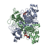
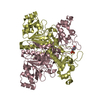
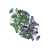
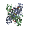
| Title | Crystal structure of Formyl-CoA Transferase (apoenzyme) from Oxalobacter formigenes | ||||||
 Components Components | Formyl-coenzyme A transferase | ||||||
 Keywords Keywords | TRANSFERASE / CoA-transferase / oxalate / oxalate degradation / intertwined / knotted fold / CAIB-BAIF family | ||||||
| Function / homology |  Function and homology information Function and homology informationformyl-CoA transferase / formyl-CoA transferase activity / oxalate catabolic process / cytoplasm Similarity search - Function | ||||||
| Biological species |  Oxalobacter formigenes (bacteria) Oxalobacter formigenes (bacteria) | ||||||
| Method |  X-RAY DIFFRACTION / SYNCHROTRON / SAD / Resolution: 2.2 Å X-RAY DIFFRACTION / SYNCHROTRON / SAD / Resolution: 2.2 Å | ||||||
 Authors Authors | Ricagno, S. / Jonsson, S. / Richards, N. / Lindqvist, Y. | ||||||
 Citation Citation | Journal: Embo J. / Year: 2003 Title: Formyl-CoA Transferase encloses the CoA binding site at the interface of an interlocked dimer Authors: Ricagno, S. / Jonsson, S. / Richards, N. / Lindqvist, Y. | ||||||
| History |
|
- Structure visualization
| Structure viewer | Molecule: MolmilJmol/JSmol |
|---|
- Downloads & links
-Download
| PDBx/mmCIF format | 1p5h.cif.gz | 182 KB | Display | PDBx/mmCIF format |
|---|---|---|---|---|
| PDB format | pdb1p5h.ent.gz | 146.5 KB | Display | PDB format |
| PDBx/mmJSON format | 1p5h.json.gz | Tree view | PDBx/mmJSON format | |
| Others |  Other downloads Other downloads |
-Validation report
| Arichive directory | https://data.pdbj.org/pub/pdb/validation_reports/p5/1p5hftp://data.pdbj.org/pub/pdb/validation_reports/p5/1p5h | HTTPS FTP |
|---|
-Related structure data


-Links
 PDBj
PDBj- Assembly
| Deposited unit | 
| ||||||||
|---|---|---|---|---|---|---|---|---|---|
| 1 |
| ||||||||
| Unit cell |
|
-Components
| #1: Protein | Mass: 47380.895 Da / Num. of mol.: 2 Source method: isolated from a genetically manipulated source Source: (gene. exp.) Oxalobacter formigenes (bacteria) / Gene: FRC / Plasmid: pET9a / Species (production host): Escherichia coli / Production host: References: UniProt: O06644, Transferases; Transferring sulfur-containing groups; CoA-transferases #2: Water | ChemComp-HOH / |  Mass: 18.015 Da / Num. of mol.: 530 / Source method: isolated from a natural source / Formula: H2O Mass: 18.015 Da / Num. of mol.: 530 / Source method: isolated from a natural source / Formula: H2O |
|---|
-Experimental details
-Experiment
| Experiment | Method: X-RAY DIFFRACTION / Number of used crystals: 2 |
|---|
- Sample preparation
| Crystal | Density Matthews: 3.01 Å3/Da / Density % sol: 59.11 % | |||||||||||||||||||||||||||||||||||
|---|---|---|---|---|---|---|---|---|---|---|---|---|---|---|---|---|---|---|---|---|---|---|---|---|---|---|---|---|---|---|---|---|---|---|---|---|
| Crystal grow | Temperature: 293 K / Method: vapor diffusion, hanging drop / pH: 7.5 Details: PEG 4000, Magnesium cloride, Hepes, pH 7.5, VAPOR DIFFUSION, HANGING DROP, temperature 293K | |||||||||||||||||||||||||||||||||||
| Crystal grow | *PLUS Temperature: 291 K / Method: vapor diffusion, hanging drop / Details: Ricagno, S., (2003) Acta Crystallogr., D59, 1276. | |||||||||||||||||||||||||||||||||||
| Components of the solutions | *PLUS
|
-Data collection
| Diffraction |
| ||||||||||||||||||
|---|---|---|---|---|---|---|---|---|---|---|---|---|---|---|---|---|---|---|---|
| Diffraction source |
| ||||||||||||||||||
| Detector |
| ||||||||||||||||||
| Radiation |
| ||||||||||||||||||
| Radiation wavelength |
| ||||||||||||||||||
| Reflection | Resolution: 2.2→25 Å / Num. all: 53823 / Num. obs: 53823 / Observed criterion σ(F): 0 / Observed criterion σ(I): 0 / Redundancy: 5.1 % / Rsym value: 0.098 / Net I/σ(I): 15.3 | ||||||||||||||||||
| Reflection shell | Resolution: 2.2→2.32 Å / Redundancy: 4.6 % / Mean I/σ(I) obs: 4.9 / Rsym value: 0.236 / % possible all: 99 | ||||||||||||||||||
| Reflection | *PLUS Num. obs: 57045 / % possible obs: 99 % / Rmerge(I) obs: 0.098 | ||||||||||||||||||
| Reflection shell | *PLUS % possible obs: 99 % / Rmerge(I) obs: 0.236 |


- Processing
| Software |
| ||||||||||||||||||||||||||||||||||||||||||||||||||||||||||||||||||||||||||||||||||||||||||||||||||||
|---|---|---|---|---|---|---|---|---|---|---|---|---|---|---|---|---|---|---|---|---|---|---|---|---|---|---|---|---|---|---|---|---|---|---|---|---|---|---|---|---|---|---|---|---|---|---|---|---|---|---|---|---|---|---|---|---|---|---|---|---|---|---|---|---|---|---|---|---|---|---|---|---|---|---|---|---|---|---|---|---|---|---|---|---|---|---|---|---|---|---|---|---|---|---|---|---|---|---|---|---|---|
| Refinement | Method to determine structure: SAD / Resolution: 2.2→24.62 Å / Cor.coef. Fo:Fc: 0.948 / Cor.coef. Fo:Fc free: 0.927 / SU B: 4.364 / SU ML: 0.113 / TLS residual ADP flag: LIKELY RESIDUAL / Cross valid method: THROUGHOUT / ESU R: 0.204 / ESU R Free: 0.171 / Stereochemistry target values: MAXIMUM LIKELIHOOD / Details: HYDROGENS HAVE BEEN ADDED IN THE RIDING POSITIONS
| ||||||||||||||||||||||||||||||||||||||||||||||||||||||||||||||||||||||||||||||||||||||||||||||||||||
| Solvent computation | Ion probe radii: 0.8 Å / Shrinkage radii: 0.8 Å / VDW probe radii: 1.4 Å / Solvent model: BABINET MODEL WITH MASK | ||||||||||||||||||||||||||||||||||||||||||||||||||||||||||||||||||||||||||||||||||||||||||||||||||||
| Displacement parameters | Biso mean: 30.915 Å2
| ||||||||||||||||||||||||||||||||||||||||||||||||||||||||||||||||||||||||||||||||||||||||||||||||||||
| Refinement step | Cycle: LAST / Resolution: 2.2→24.62 Å
| ||||||||||||||||||||||||||||||||||||||||||||||||||||||||||||||||||||||||||||||||||||||||||||||||||||
| Refine LS restraints |
| ||||||||||||||||||||||||||||||||||||||||||||||||||||||||||||||||||||||||||||||||||||||||||||||||||||
| LS refinement shell | Resolution: 2.2→2.257 Å / Total num. of bins used: 20 /
| ||||||||||||||||||||||||||||||||||||||||||||||||||||||||||||||||||||||||||||||||||||||||||||||||||||
| Refinement TLS params. | Method: refined / Refine-ID: X-RAY DIFFRACTION
| ||||||||||||||||||||||||||||||||||||||||||||||||||||||||||||||||||||||||||||||||||||||||||||||||||||
| Refinement TLS group |
| ||||||||||||||||||||||||||||||||||||||||||||||||||||||||||||||||||||||||||||||||||||||||||||||||||||
| Refinement | *PLUS Highest resolution: 2.2 Å / Lowest resolution: 25 Å / Rfactor Rfree: 0.209 / Rfactor Rwork: 0.171 | ||||||||||||||||||||||||||||||||||||||||||||||||||||||||||||||||||||||||||||||||||||||||||||||||||||
| Solvent computation | *PLUS | ||||||||||||||||||||||||||||||||||||||||||||||||||||||||||||||||||||||||||||||||||||||||||||||||||||
| Displacement parameters | *PLUS | ||||||||||||||||||||||||||||||||||||||||||||||||||||||||||||||||||||||||||||||||||||||||||||||||||||
| LS refinement shell | *PLUS Rfactor Rfree: 0.219 / Rfactor Rwork: 0.19 |
