Protocol: SINGLE WAVELENGTH / Monochromatic (M) / Laue (L): M / Scattering type: x-ray
Radiation wavelength
Wavelength: 1 Å / Relative weight: 1
Reflection
Resolution: 2.28→49.55 Å / Num. obs: 65614 / % possible obs: 85.6 % / Observed criterion σ(I): 2.13 / Redundancy: 8.2 % / Biso Wilson estimate: 34.35 Å2 / Rmerge(I) obs: 0.05 / Net I/σ(I): 37.12
Reflection shell
Resolution: 2.28→2.36 Å / Redundancy: 5.8 % / Rmerge(I) obs: 0.2 / Mean I/σ(I) obs: 4.19 / % possible all: 72.9
-
Processing
Software
Name
Version
Classification
PHENIX
(PHENIX.REFINE)
refinement
HKL-2000
datareduction
HKL-2000
datascaling
PHASER
phasing
Refinement
Method to determine structure: MOLECULAR REPLACEMENT / Resolution: 2.28→49.554 Å / SU ML: 0.28 / σ(F): 2.13 / Phase error: 23.23 / Stereochemistry target values: ML Details: IN BOTH CHAIN A AND B, THE RESIDUES FROM 656-659 AND 671-678 REGIONS ARE DISORDERED
Rfactor
Num. reflection
% reflection
Rfree
0.2379
3087
5.1 %
Rwork
0.1847
-
-
obs
0.1874
60614
85.57 %
Solvent computation
Shrinkage radii: 0.9 Å / VDW probe radii: 1.11 Å / Solvent model: FLAT BULK SOLVENT MODEL / Bsol: 42.207 Å2 / ksol: 0.347 e/Å3
Displacement parameters
Biso mean: 4.54 Å2
Baniso -1
Baniso -2
Baniso -3
1-
12.6395 Å2
0 Å2
0 Å2
2-
-
-8.0802 Å2
0 Å2
3-
-
-
-4.5593 Å2
Refinement step
Cycle: LAST / Resolution: 2.28→49.554 Å
Protein
Nucleic acid
Ligand
Solvent
Total
Num. atoms
11474
0
0
298
11772
Refine LS restraints
Refine-ID
Type
Dev ideal
Number
X-RAY DIFFRACTION
f_bond_d
0.012
12256
X-RAY DIFFRACTION
f_angle_d
1.116
16085
X-RAY DIFFRACTION
f_dihedral_angle_d
15.383
4479
X-RAY DIFFRACTION
f_chiral_restr
0.077
1663
X-RAY DIFFRACTION
f_plane_restr
0.004
2047
LS refinement shell
Resolution (Å)
Rfactor Rfree
Num. reflection Rfree
Rfactor Rwork
Num. reflection Rwork
Refine-ID
% reflection obs (%)
2.28-2.3615
0.2981
272
0.2235
4820
X-RAY DIFFRACTION
73
2.3615-2.456
0.3029
283
0.2072
4919
X-RAY DIFFRACTION
74
2.456-2.5678
0.2523
292
0.193
4950
X-RAY DIFFRACTION
75
2.5678-2.7032
0.2895
276
0.1973
5162
X-RAY DIFFRACTION
78
2.7032-2.8725
0.2585
280
0.1938
5415
X-RAY DIFFRACTION
81
2.8725-3.0943
0.2596
284
0.1967
5813
X-RAY DIFFRACTION
86
3.0943-3.4056
0.2426
335
0.1891
6215
X-RAY DIFFRACTION
93
3.4056-3.8982
0.2434
327
0.1776
6553
X-RAY DIFFRACTION
97
3.8982-4.9107
0.2102
344
0.1561
6698
X-RAY DIFFRACTION
98
4.9107-49.5661
0.2123
394
0.1946
6982
X-RAY DIFFRACTION
99
+
About Yorodumi
-
News
-
Feb 9, 2022. New format data for meta-information of EMDB entries
New format data for meta-information of EMDB entries
Version 3 of the EMDB header file is now the official format.
The previous official version 1.9 will be removed from the archive.
In the structure databanks used in Yorodumi, some data are registered as the other names, "COVID-19 virus" and "2019-nCoV". Here are the details of the virus and the list of structure data.
Jan 31, 2019. EMDB accession codes are about to change! (news from PDBe EMDB page)
EMDB accession codes are about to change! (news from PDBe EMDB page)
The allocation of 4 digits for EMDB accession codes will soon come to an end. Whilst these codes will remain in use, new EMDB accession codes will include an additional digit and will expand incrementally as the available range of codes is exhausted. The current 4-digit format prefixed with “EMD-” (i.e. EMD-XXXX) will advance to a 5-digit format (i.e. EMD-XXXXX), and so on. It is currently estimated that the 4-digit codes will be depleted around Spring 2019, at which point the 5-digit format will come into force.
The EM Navigator/Yorodumi systems omit the EMD- prefix.
Related info.:Q: What is EMD? / ID/Accession-code notation in Yorodumi/EM Navigator
Yorodumi is a browser for structure data from EMDB, PDB, SASBDB, etc.
This page is also the successor to EM Navigator detail page, and also detail information page/front-end page for Omokage search.
The word "yorodu" (or yorozu) is an old Japanese word meaning "ten thousand". "mi" (miru) is to see.
Related info.:EMDB / PDB / SASBDB / Comparison of 3 databanks / Yorodumi Search / Aug 31, 2016. New EM Navigator & Yorodumi / Yorodumi Papers / Jmol/JSmol / Function and homology information / Changes in new EM Navigator and Yorodumi
 Movie
Movie Controller


 Open data
 Basic information
 Components
 Keywords
 Function and homology information
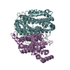
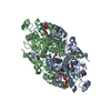
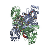
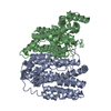
 STAPHYLOTHERMUS MARINUS (archaea)
 X-RAY DIFFRACTION /
  Authors
 Citation
 Structure visualization
 Downloads & links
 Other downloads


 PDBj
PDBj

 Assembly


 Mass: 18.015 Da / Num. of mol.: 298 / Source method: isolated from a natural source / Formula: H2O
 Sample preparation
 / Beamline: 6C1 / Wavelength: 1
  Processing