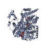
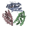
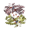
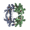
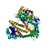
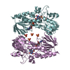
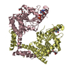
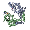
A: METHYLMALONIC ACIDURIA TYPE A PROTEIN, MITOCHONDRIAL B: METHYLMALONIC ACIDURIA TYPE A PROTEIN, MITOCHONDRIAL C: METHYLMALONIC ACIDURIA TYPE A PROTEIN, MITOCHONDRIAL D: METHYLMALONIC ACIDURIA TYPE A PROTEIN, MITOCHONDRIAL hetero molecules
Protocol: SINGLE WAVELENGTH / Monochromatic (M) / Laue (L): M / Scattering type: x-ray
Radiation wavelength
Wavelength: 0.9763 Å / Relative weight: 1
Reflection
Resolution: 2.64→27.43 Å / Num. obs: 45149 / % possible obs: 99.8 % / Observed criterion σ(I): 2 / Redundancy: 4 % / Biso Wilson estimate: 82.56 Å2 / Rmerge(I) obs: 0.07 / Net I/σ(I): 10.2
Reflection shell
Resolution: 2.64→2.78 Å / Redundancy: 3.8 % / Rmerge(I) obs: 0.61 / Mean I/σ(I) obs: 2 / % possible all: 100
-
Processing
Software
Name
Version
Classification
BUSTER
2.8.0
refinement
iMOSFLM
datareduction
SCALA
datascaling
PHASER
phasing
Refinement
Method to determine structure: MOLECULAR REPLACEMENT / Resolution: 2.64→27.43 Å / Cor.coef. Fo:Fc: 0.9438 / Cor.coef. Fo:Fc free: 0.9249 / Cross valid method: THROUGHOUT / σ(F): 0 Details: THE PROTEIN WAS CRYSTALLIZED PHOSPHOAMINOPHOSPHONIC ACID-GUANYLATE ESTER BUT GDP MOLECULES WERE IDENTIFIED BASED ON ELECTRON DENSITY
Rfactor
Num. reflection
% reflection
Selection details
Rfree
0.2219
2272
5.05 %
RANDOM
Rwork
0.1819
-
-
-
obs
0.184
45022
-
-
Displacement parameters
Biso mean: 84.87 Å2
Baniso -1
Baniso -2
Baniso -3
1-
-6.6144 Å2
0 Å2
0 Å2
2-
-
-6.6144 Å2
0 Å2
3-
-
-
13.2288 Å2
Refine analyze
Luzzati coordinate error obs: 0.364 Å
Refinement step
Cycle: LAST / Resolution: 2.64→27.43 Å
Protein
Nucleic acid
Ligand
Solvent
Total
Num. atoms
8918
0
155
87
9160
Refine LS restraints
Refine-ID
Type
Dev ideal
Number
Restraint function
Weight
X-RAY DIFFRACTION
t_bond_d
0.013
9218
HARMONIC
2
X-RAY DIFFRACTION
t_angle_deg
1.35
12501
HARMONIC
2
X-RAY DIFFRACTION
t_dihedral_angle_d
3068
SINUSOIDAL
2
X-RAY DIFFRACTION
t_incorr_chiral_ct
X-RAY DIFFRACTION
t_pseud_angle
X-RAY DIFFRACTION
t_trig_c_planes
182
HARMONIC
2
X-RAY DIFFRACTION
t_gen_planes
1328
HARMONIC
5
X-RAY DIFFRACTION
t_it
9058
HARMONIC
20
X-RAY DIFFRACTION
t_nbd
X-RAY DIFFRACTION
t_omega_torsion
3.14
X-RAY DIFFRACTION
t_other_torsion
19.57
X-RAY DIFFRACTION
t_improper_torsion
X-RAY DIFFRACTION
t_chiral_improper_torsion
1256
SEMIHARMONIC
5
X-RAY DIFFRACTION
t_sum_occupancies
X-RAY DIFFRACTION
t_utility_distance
X-RAY DIFFRACTION
t_utility_angle
X-RAY DIFFRACTION
t_utility_torsion
X-RAY DIFFRACTION
t_ideal_dist_contact
9962
SEMIHARMONIC
4
LS refinement shell
Resolution: 2.64→2.71 Å / Total num. of bins used: 20
Rfactor
Num. reflection
% reflection
Rfree
0.2774
150
4.55 %
Rwork
0.2162
3146
-
all
0.219
3296
-
Refinement TLS params.
Method: refined / Refine-ID: X-RAY DIFFRACTION
ID
L11 (°2)
L12 (°2)
L13 (°2)
L22 (°2)
L23 (°2)
L33 (°2)
S11 (Å °)
S12 (Å °)
S13 (Å °)
S21 (Å °)
S22 (Å °)
S23 (Å °)
S31 (Å °)
S32 (Å °)
S33 (Å °)
T11 (Å2)
T12 (Å2)
T13 (Å2)
T22 (Å2)
T23 (Å2)
T33 (Å2)
Origin x (Å)
Origin y (Å)
Origin z (Å)
1
0.7874
0.3415
-0.3026
2.0901
-0.5705
0.4671
-0.1856
-0.0024
-0.0605
0.2775
0.337
0.2363
-0.0795
-0.2199
-0.1514
-0.0333
0.1043
0.0163
0.063
0.1297
0.0297
29.0325
77.303
101.049
2
1.7143
-0.9302
-2.008
2.0798
0.6082
2.9377
-0.5041
0.0823
-0.0456
-0.1035
0.358
-0.1655
1.024
0.0196
0.146
0.3937
0.0182
-0.0189
0.0959
-0.0365
-0.0308
46.651
76.2402
76.8144
3
0.0528
0.6453
0.8183
1.9082
1.3876
1.5458
-0.2318
0.1801
-0.1764
-0.1628
0.4482
-0.2633
-0.0237
0.755
-0.2164
-0.0439
-0.0493
0.0869
0.4131
-0.0453
0.0242
44.564
4.4774
98.4405
4
1.2372
-0.3871
0.5589
1.0963
-0.3252
1.585
-0.3069
-0.253
-0.057
-0.0007
0.2539
0.0414
-0.0444
-0.1629
0.0531
-0.0425
0.075
0.0281
-0.0381
0.0393
-0.1117
27.4595
3.0494
73.5652
Refinement TLS group
ID
Refine-ID
Refine TLS-ID
Selection details
1
X-RAY DIFFRACTION
1
(CHAINA)
2
X-RAY DIFFRACTION
2
(CHAINB)
3
X-RAY DIFFRACTION
3
(CHAINC)
4
X-RAY DIFFRACTION
4
(CHAIND)
+
About Yorodumi
-
News
-
Feb 9, 2022. New format data for meta-information of EMDB entries
New format data for meta-information of EMDB entries
Version 3 of the EMDB header file is now the official format.
The previous official version 1.9 will be removed from the archive.
In the structure databanks used in Yorodumi, some data are registered as the other names, "COVID-19 virus" and "2019-nCoV". Here are the details of the virus and the list of structure data.
Jan 31, 2019. EMDB accession codes are about to change! (news from PDBe EMDB page)
EMDB accession codes are about to change! (news from PDBe EMDB page)
The allocation of 4 digits for EMDB accession codes will soon come to an end. Whilst these codes will remain in use, new EMDB accession codes will include an additional digit and will expand incrementally as the available range of codes is exhausted. The current 4-digit format prefixed with “EMD-” (i.e. EMD-XXXX) will advance to a 5-digit format (i.e. EMD-XXXXX), and so on. It is currently estimated that the 4-digit codes will be depleted around Spring 2019, at which point the 5-digit format will come into force.
The EM Navigator/Yorodumi systems omit the EMD- prefix.
Related info.:Q: What is EMD? / ID/Accession-code notation in Yorodumi/EM Navigator
Yorodumi is a browser for structure data from EMDB, PDB, SASBDB, etc.
This page is also the successor to EM Navigator detail page, and also detail information page/front-end page for Omokage search.
The word "yorodu" (or yorozu) is an old Japanese word meaning "ten thousand". "mi" (miru) is to see.
Related info.:EMDB / PDB / SASBDB / Comparison of 3 databanks / Yorodumi Search / Aug 31, 2016. New EM Navigator & Yorodumi / Yorodumi Papers / Jmol/JSmol / Function and homology information / Changes in new EM Navigator and Yorodumi
 Movie
Movie Controller


 Open data
 Basic information
 Components
 Keywords
 Function and homology information
 HOMO SAPIENS (human)
 X-RAY DIFFRACTION /
  Authors
 Citation
 Structure visualization
 Downloads & links
 Other downloads
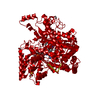

 PDBj
PDBj Assembly
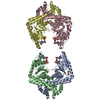


 Mass: 414.488 Da / Num. of mol.: 3 / Source method: obtained synthetically / Formula: C18H38O10 / Comment: precipitant*YM
 Type: RNA linking / Mass: 443.201 Da / Num. of mol.: 4 / Source method: obtained synthetically / Formula: C10H15N5O11P2 / Comment: GDP, energy-carrying molecule*YM
 Mass: 18.015 Da / Num. of mol.: 87 / Source method: isolated from a natural source / Formula: H2O
 Sample preparation
 / Beamline: I03 / Wavelength: 0.9763
  Processing