- PDB-3svm: Human MPP8 - human DNMT3AK47me2 peptide -
+
Open data
ID or keywords:
Loading...
-
Basic information
Entry
Database: PDB / ID: 3svm
Title
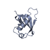
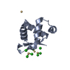
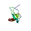
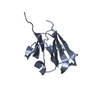
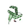
Human MPP8 - human DNMT3AK47me2 peptide
Components
DNA (cytosine-5)-methyltransferase 3A
M-phase phosphoprotein 8
Keywords
TRANSFERASE / epigenetics / methyl-lysine binding / Chromodomain / The dimethylated human DNMT3AK47me2 is recognized by the chromodomain of MPP8 / MPP8 chromodomain / dimethylated lysine
Function / homology
Function and homology information
HUSH2 complex / HUSH complex / constitutive heterochromatin formation / transposable element silencing by heterochromatin formation / transposable element silencing by piRNA-mediated DNA methylation / protein-cysteine methyltransferase activity / positive regulation of cellular response to hypoxia / cellular response to bisphenol A / regulatory ncRNA-mediated heterochromatin formation / unmethylated CpG binding ...HUSH2 complex / HUSH complex / constitutive heterochromatin formation / transposable element silencing by heterochromatin formation / transposable element silencing by piRNA-mediated DNA methylation / protein-cysteine methyltransferase activity / positive regulation of cellular response to hypoxia / cellular response to bisphenol A / regulatory ncRNA-mediated heterochromatin formation / unmethylated CpG binding / DNA (cytosine-5-)-methyltransferase / DNA (cytosine-5-)-methyltransferase activity / autosome genomic imprinting / SUMOylation of DNA methylation proteins / XY body / oocyte development / response to vitamin A / histone H3K9me2/3 reader activity / DNA methylation-dependent constitutive heterochromatin formation / negative regulation of type I interferon production / response to ionizing radiation / hepatocyte apoptotic process / negative regulation of gene expression via chromosomal CpG island methylation / negative regulation of gene expression, epigenetic / lncRNA binding / cellular response to ethanol / chromosome, centromeric region / catalytic complex / heterochromatin / Transferases; Transferring one-carbon groups; Methyltransferases / Regulation of endogenous retroelements by the Human Silencing Hub (HUSH) complex / DNA methylation / post-embryonic development / PRC2 methylates histones and DNA / Defective pyroptosis / response to cocaine / cellular response to amino acid stimulus / Regulation of endogenous retroelements by Piwi-interacting RNAs (piRNAs) / euchromatin / Negative Regulation of CDH1 Gene Transcription / response to lead ion / response to toxic substance / neuron differentiation / nuclear matrix / RMTs methylate histone arginines / transcription corepressor activity / response to estradiol / nucleosome / methylation / spermatogenesis / cellular response to hypoxia / RNA polymerase II-specific DNA-binding transcription factor binding / RNA polymerase II cis-regulatory region sequence-specific DNA binding / response to xenobiotic stimulus / negative regulation of DNA-templated transcription / chromatin binding / chromatin / nucleolus / negative regulation of transcription by RNA polymerase II / DNA binding / zinc ion binding / nucleoplasm / identical protein binding / nucleus / plasma membrane / cytoplasm / cytosol Similarity search - Function
In the structure databanks used in Yorodumi, some data are registered as the other names, "COVID-19 virus" and "2019-nCoV". Here are the details of the virus and the list of structure data.
Jan 31, 2019. EMDB accession codes are about to change! (news from PDBe EMDB page)
EMDB accession codes are about to change! (news from PDBe EMDB page)
The allocation of 4 digits for EMDB accession codes will soon come to an end. Whilst these codes will remain in use, new EMDB accession codes will include an additional digit and will expand incrementally as the available range of codes is exhausted. The current 4-digit format prefixed with “EMD-” (i.e. EMD-XXXX) will advance to a 5-digit format (i.e. EMD-XXXXX), and so on. It is currently estimated that the 4-digit codes will be depleted around Spring 2019, at which point the 5-digit format will come into force.
The EM Navigator/Yorodumi systems omit the EMD- prefix.
Related info.:Q: What is EMD? / ID/Accession-code notation in Yorodumi/EM Navigator
Yorodumi is a browser for structure data from EMDB, PDB, SASBDB, etc.
This page is also the successor to EM Navigator detail page, and also detail information page/front-end page for Omokage search.
The word "yorodu" (or yorozu) is an old Japanese word meaning "ten thousand". "mi" (miru) is to see.
Related info.:EMDB / PDB / SASBDB / Comparison of 3 databanks / Yorodumi Search / Aug 31, 2016. New EM Navigator & Yorodumi / Yorodumi Papers / Jmol/JSmol / Function and homology information / Changes in new EM Navigator and Yorodumi
 Movie
Movie Controller


 Open data
 Basic information
 Components
 Keywords
 Function and homology information
 Homo sapiens (human)
 X-RAY DIFFRACTION /
  Authors
 Citation
 Structure visualization
 Downloads & links
 Other downloads


 PDBj
PDBj


 Assembly
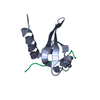


 Mass: 18.015 Da / Num. of mol.: 24 / Source method: isolated from a natural source / Formula: H2O
 Sample preparation
 / Beamline: 22-ID / Wavelength: 1 Å
 Processing