- PDB-1zzk: Crystal Structure of the third KH domain of hnRNP K at 0.95A reso... -
+
Open data
ID or keywords:
Loading...
-
Basic information
Entry
Database: PDB / ID: 1zzk
Title
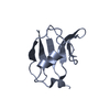
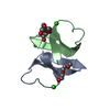
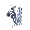
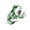
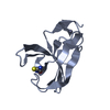
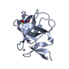
Crystal Structure of the third KH domain of hnRNP K at 0.95A resolution
Components
Heterogeneous nuclear ribonucleoprotein K
Keywords
DNA BINDING PROTEIN / KH domian / alpha-beta fold
Function / homology
Function and homology information
regulation of intrinsic apoptotic signaling pathway in response to DNA damage by p53 class mediator / positive regulation of low-density lipoprotein particle clearance / random inactivation of X chromosome / regulatory ncRNA-mediated heterochromatin formation / regulation of mRNA splicing, via spliceosome / SUMOylation of RNA binding proteins / podosome / Processing of Capped Intron-Containing Pre-mRNA / negative regulation of mRNA splicing, via spliceosome / RNA processing ...regulation of intrinsic apoptotic signaling pathway in response to DNA damage by p53 class mediator / positive regulation of low-density lipoprotein particle clearance / random inactivation of X chromosome / regulatory ncRNA-mediated heterochromatin formation / regulation of mRNA splicing, via spliceosome / SUMOylation of RNA binding proteins / podosome / Processing of Capped Intron-Containing Pre-mRNA / negative regulation of mRNA splicing, via spliceosome / RNA processing / catalytic step 2 spliceosome / mRNA Splicing - Major Pathway / HCMV Late Events / mRNA splicing, via spliceosome / cytoplasmic stress granule / cadherin binding / ribonucleoprotein complex / protein domain specific binding / focal adhesion / negative regulation of DNA-templated transcription / mRNA binding / regulation of transcription by RNA polymerase II / negative regulation of apoptotic process / chromatin / signal transduction / positive regulation of transcription by RNA polymerase II / DNA binding / RNA binding / extracellular exosome / nucleoplasm / membrane / identical protein binding / nucleus / cytoplasm Similarity search - Function
ROK, N-terminal / ROKNT (NUC014) domain / K Homology domain, type 1 / KH domain / K Homology domain, type 1 / Ribosomal Protein S8; Chain: A, domain 1 / Type-1 KH domain profile. / K Homology domain, type 1 superfamily / K Homology domain / K homology RNA-binding domain ...ROK, N-terminal / ROKNT (NUC014) domain / K Homology domain, type 1 / KH domain / K Homology domain, type 1 / Ribosomal Protein S8; Chain: A, domain 1 / Type-1 KH domain profile. / K Homology domain, type 1 superfamily / K Homology domain / K homology RNA-binding domain / 2-Layer Sandwich / Alpha Beta Similarity search - Domain/homology
In the structure databanks used in Yorodumi, some data are registered as the other names, "COVID-19 virus" and "2019-nCoV". Here are the details of the virus and the list of structure data.
Jan 31, 2019. EMDB accession codes are about to change! (news from PDBe EMDB page)
EMDB accession codes are about to change! (news from PDBe EMDB page)
The allocation of 4 digits for EMDB accession codes will soon come to an end. Whilst these codes will remain in use, new EMDB accession codes will include an additional digit and will expand incrementally as the available range of codes is exhausted. The current 4-digit format prefixed with “EMD-” (i.e. EMD-XXXX) will advance to a 5-digit format (i.e. EMD-XXXXX), and so on. It is currently estimated that the 4-digit codes will be depleted around Spring 2019, at which point the 5-digit format will come into force.
The EM Navigator/Yorodumi systems omit the EMD- prefix.
Related info.:Q: What is EMD? / ID/Accession-code notation in Yorodumi/EM Navigator
Yorodumi is a browser for structure data from EMDB, PDB, SASBDB, etc.
This page is also the successor to EM Navigator detail page, and also detail information page/front-end page for Omokage search.
The word "yorodu" (or yorozu) is an old Japanese word meaning "ten thousand". "mi" (miru) is to see.
Related info.:EMDB / PDB / SASBDB / Comparison of 3 databanks / Yorodumi Search / Aug 31, 2016. New EM Navigator & Yorodumi / Yorodumi Papers / Jmol/JSmol / Function and homology information / Changes in new EM Navigator and Yorodumi
 Movie
Movie Controller

 Yorodumi
 Open data
 Basic information
 Components
 Keywords
 Function and homology information
 Homo sapiens (human)
 X-RAY DIFFRACTION /
  Authors
 Citation
 Structure visualization
 Downloads & links
 Other downloads


 PDBj
PDBj


 Assembly

 Mass: 18.015 Da / Num. of mol.: 58 / Source method: isolated from a natural source / Formula: H2O
 Sample preparation
 / Beamline: ID14-2 / Wavelength: 0.933 Å
 Processing