 Movie
Movie Controller

[English] 日本語
 Yorodumi
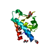
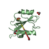
- PDB-4oeo: High resolution crystal structure of the unliganded ZO-1 PDZ1 domain -
+ Open data

- Basic information
| Entry | Database: PDB / ID: 4oeo | ||||||
|---|---|---|---|---|---|---|---|
| Title | High resolution crystal structure of the unliganded ZO-1 PDZ1 domain | ||||||
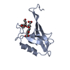
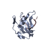
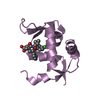
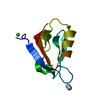
 Components Components | Tight junction protein ZO-1 | ||||||
 Keywords Keywords | CELL ADHESION / MAGUK / PDZ1 / SCAFFOLDING / Claudin / tight junction assembly | ||||||
| Function / homology |  Function and homology information Function and homology informationpositive regulation of blood-brain barrier permeability / ameloblast differentiation / adherens junction maintenance / RUNX1 regulates expression of components of tight junctions / positive regulation of cell-cell adhesion mediated by cadherin / establishment of endothelial intestinal barrier / SARS-CoV-2 targets PDZ proteins in cell-cell junction / protein localization to cell-cell junction / regulation of cell junction assembly / Regulation of gap junction activity ...positive regulation of blood-brain barrier permeability / ameloblast differentiation / adherens junction maintenance / RUNX1 regulates expression of components of tight junctions / positive regulation of cell-cell adhesion mediated by cadherin / establishment of endothelial intestinal barrier / SARS-CoV-2 targets PDZ proteins in cell-cell junction / protein localization to cell-cell junction / regulation of cell junction assembly / Regulation of gap junction activity / protein localization to bicellular tight junction / cell-cell junction organization / protein localization to adherens junction / gap junction / Apoptotic cleavage of cell adhesion proteins / negative regulation of stress fiber assembly / Signaling by Hippo / actomyosin structure organization / cell-cell junction assembly / podosome / tight junction / regulation of bicellular tight junction assembly / apical junction complex / positive regulation of sprouting angiogenesis / regulation of cytoskeleton organization / maintenance of blood-brain barrier / Dengue Virus-Host Interactions / bicellular tight junction / cell adhesion molecule binding / adherens junction / cell-cell adhesion / apical part of cell / cell junction / actin cytoskeleton organization / basolateral plasma membrane / calmodulin binding / positive regulation of cell migration / cadherin binding / positive regulation of cell population proliferation / negative regulation of apoptotic process / protein-containing complex / plasma membrane / cytosol / cytoplasm Similarity search - Function | ||||||
| Biological species |  Homo sapiens (human) Homo sapiens (human) | ||||||
| Method |  X-RAY DIFFRACTION / SYNCHROTRON / MOLECULAR REPLACEMENT / Resolution: 1.9 Å X-RAY DIFFRACTION / SYNCHROTRON / MOLECULAR REPLACEMENT / Resolution: 1.9 Å | ||||||
 Authors Authors | Nomme, J. / Lavie, A. | ||||||
 Citation Citation | Journal: J.Biol.Chem. / Year: 2015 Title: Structural Basis of a Key Factor Regulating the Affinity between the Zonula Occludens First PDZ Domain and Claudins. Authors: Nomme, J. / Antanasijevic, A. / Caffrey, M. / Van Itallie, C.M. / Anderson, J.M. / Fanning, A.S. / Lavie, A. | ||||||
| History |
|
- Structure visualization
| Structure viewer | Molecule: MolmilJmol/JSmol |
|---|
- Downloads & links
-Download
| PDBx/mmCIF format | 4oeo.cif.gz | 70 KB | Display | PDBx/mmCIF format |
|---|---|---|---|---|
| PDB format | pdb4oeo.ent.gz | 51.3 KB | Display | PDB format |
| PDBx/mmJSON format | 4oeo.json.gz | Tree view | PDBx/mmJSON format | |
| Others |  Other downloads Other downloads |
-Validation report
| Arichive directory | https://data.pdbj.org/pub/pdb/validation_reports/oe/4oeoftp://data.pdbj.org/pub/pdb/validation_reports/oe/4oeo | HTTPS FTP |
|---|
-Related structure data


-Links
 PDBj
PDBj


- Assembly
| Deposited unit | 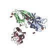
| ||||||||
|---|---|---|---|---|---|---|---|---|---|
| 1 | 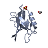
| ||||||||
| 2 | 
| ||||||||
| 3 | 
| ||||||||
| 4 | 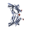
| ||||||||
| 5 | 
| ||||||||
| Unit cell |
| ||||||||
| Components on special symmetry positions |
|
-Components
| #1: Protein | Mass: 11486.908 Da / Num. of mol.: 3 / Fragment: PDZ1 domain, UNP residues 18-110 Source method: isolated from a genetically manipulated source Source: (gene. exp.) Homo sapiens (human) / Gene: TJP1, ZO1 / Plasmid: pET14b / Production host:  #2: Chemical |   Mass: 59.044 Da / Num. of mol.: 3 / Source method: obtained synthetically / Formula: C2H3O2 Mass: 59.044 Da / Num. of mol.: 3 / Source method: obtained synthetically / Formula: C2H3O2#3: Chemical |   Mass: 96.063 Da / Num. of mol.: 2 / Source method: obtained synthetically / Formula: SO4 Mass: 96.063 Da / Num. of mol.: 2 / Source method: obtained synthetically / Formula: SO4#4: Chemical |   Mass: 546.646 Da / Num. of mol.: 2 / Source method: obtained synthetically / Formula: C24H50O13 / Comment: precipitant*YM Mass: 546.646 Da / Num. of mol.: 2 / Source method: obtained synthetically / Formula: C24H50O13 / Comment: precipitant*YM#5: Water | ChemComp-HOH / |  Mass: 18.015 Da / Num. of mol.: 96 / Source method: isolated from a natural source / Formula: H2O Mass: 18.015 Da / Num. of mol.: 96 / Source method: isolated from a natural source / Formula: H2O |
|---|
-Experimental details
-Experiment
| Experiment | Method: X-RAY DIFFRACTION / Number of used crystals: 1 |
|---|
- Sample preparation
| Crystal | Density Matthews: 2.37 Å3/Da / Density % sol: 48.07 % |
|---|---|
| Crystal grow | Temperature: 293 K / Method: vapor diffusion, hanging drop / pH: 4.6 Details: 30% PEG2K MME, 0.1 M NaAc pH4.6, 0.2 M AmSO4, VAPOR DIFFUSION, HANGING DROP, temperature 293K |
-Data collection
| Diffraction | Mean temperature: 100 K |
|---|---|
| Diffraction source | Source: SYNCHROTRON / Site: APS  / Beamline: 21-ID-G / Wavelength: 0.97856 Å / Beamline: 21-ID-G / Wavelength: 0.97856 Å |
| Detector | Type: MARMOSAIC 300 mm CCD / Detector: CCD / Date: Jul 31, 2013 |
| Radiation | Monochromator: c111 / Protocol: SINGLE WAVELENGTH / Monochromatic (M) / Laue (L): M / Scattering type: x-ray |
| Radiation wavelength | Wavelength: 0.97856 Å / Relative weight: 1 |
| Reflection | Resolution: 1.89→30 Å / Num. all: 24585 / Num. obs: 24585 / % possible obs: 95.1 % / Observed criterion σ(F): 0 / Observed criterion σ(I): 0 / Rsym value: 0.028 / Net I/σ(I): 17.46 |
| Reflection shell | Resolution: 1.89→2.01 Å / Mean I/σ(I) obs: 2.71 / Num. unique all: 3900 / Rsym value: 0.448 / % possible all: 94.2 |
- Processing
| Software |
| |||||||||||||||||||||||||||||||||||||||||||||||||||||||||||||||||||||||||||||||||||||||||||||||||||||||||
|---|---|---|---|---|---|---|---|---|---|---|---|---|---|---|---|---|---|---|---|---|---|---|---|---|---|---|---|---|---|---|---|---|---|---|---|---|---|---|---|---|---|---|---|---|---|---|---|---|---|---|---|---|---|---|---|---|---|---|---|---|---|---|---|---|---|---|---|---|---|---|---|---|---|---|---|---|---|---|---|---|---|---|---|---|---|---|---|---|---|---|---|---|---|---|---|---|---|---|---|---|---|---|---|---|---|---|
| Refinement | Method to determine structure: MOLECULAR REPLACEMENT / Resolution: 1.9→30 Å / Cor.coef. Fo:Fc: 0.963 / Cor.coef. Fo:Fc free: 0.941 / SU B: 5.248 / SU ML: 0.143 / Cross valid method: THROUGHOUT / σ(F): 0 / ESU R: 0.169 / ESU R Free: 0.159 / Stereochemistry target values: MAXIMUM LIKELIHOOD / Details: HYDROGENS HAVE BEEN ADDED IN THE RIDING POSITIONS
| |||||||||||||||||||||||||||||||||||||||||||||||||||||||||||||||||||||||||||||||||||||||||||||||||||||||||
| Solvent computation | Ion probe radii: 0.8 Å / Shrinkage radii: 0.8 Å / VDW probe radii: 1.2 Å / Solvent model: MASK | |||||||||||||||||||||||||||||||||||||||||||||||||||||||||||||||||||||||||||||||||||||||||||||||||||||||||
| Displacement parameters | Biso mean: 36.904 Å2
| |||||||||||||||||||||||||||||||||||||||||||||||||||||||||||||||||||||||||||||||||||||||||||||||||||||||||
| Refinement step | Cycle: LAST / Resolution: 1.9→30 Å
| |||||||||||||||||||||||||||||||||||||||||||||||||||||||||||||||||||||||||||||||||||||||||||||||||||||||||
| Refine LS restraints |
| |||||||||||||||||||||||||||||||||||||||||||||||||||||||||||||||||||||||||||||||||||||||||||||||||||||||||
| LS refinement shell | Resolution: 1.9→1.949 Å / Total num. of bins used: 20
|
