 Movie
Movie Controller

[English] 日本語
 Yorodumi
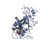
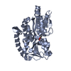
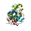
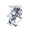
- PDB-3swc: GLP (G9a-like protein) SET domain in complex with Dnmt3aK44me2 peptide -
+ Open data

- Basic information
| Entry | Database: PDB / ID: 3swc | ||||||
|---|---|---|---|---|---|---|---|
| Title | GLP (G9a-like protein) SET domain in complex with Dnmt3aK44me2 peptide | ||||||
 Components Components |
| ||||||
 Keywords Keywords | TRANSFERASE / epigenetics / non-histone lysine methylation / SET domain / protein lysine methyltransferase | ||||||
| Function / homology |  Function and homology information Function and homology informationDNA (cytosine-5-)-methyltransferase activity, acting on CpN substrates / negative regulation of white fat cell differentiation / epigenetic programming of gene expression / PRC2 methylates histones and DNA / histone H3K9me2 methyltransferase activity / histone H3K9 trimethyltransferase activity / [histone H3]-lysine9 N-methyltransferase / peptidyl-lysine monomethylation / peptidyl-lysine dimethylation / histone H3K9 methyltransferase activity ...DNA (cytosine-5-)-methyltransferase activity, acting on CpN substrates / negative regulation of white fat cell differentiation / epigenetic programming of gene expression / PRC2 methylates histones and DNA / histone H3K9me2 methyltransferase activity / histone H3K9 trimethyltransferase activity / [histone H3]-lysine9 N-methyltransferase / peptidyl-lysine monomethylation / peptidyl-lysine dimethylation / histone H3K9 methyltransferase activity / histone H3K9 monomethyltransferase activity / histone H3K27 methyltransferase activity / transposable element silencing by piRNA-mediated DNA methylation / RMTs methylate histone arginines / protein-cysteine methyltransferase activity / positive regulation of cellular response to hypoxia / cellular response to bisphenol A / regulatory ncRNA-mediated heterochromatin formation / facultative heterochromatin formation / unmethylated CpG binding / DNA (cytosine-5-)-methyltransferase / genomic imprinting / DNA (cytosine-5-)-methyltransferase activity / autosome genomic imprinting / C2H2 zinc finger domain binding / protein-lysine N-methyltransferase activity / XY body / response to vitamin A / beige fat cell differentiation / DNA methylation-dependent constitutive heterochromatin formation / response to ionizing radiation / hepatocyte apoptotic process / negative regulation of gene expression via chromosomal CpG island methylation / lncRNA binding / Transcriptional Regulation by E2F6 / Transcriptional Regulation by VENTX / regulation of embryonic development / cellular response to ethanol / chromosome, centromeric region / catalytic complex / response to fungicide / heterochromatin / epigenetic regulation of gene expression / brown fat cell differentiation / Transferases; Transferring one-carbon groups; Methyltransferases / Regulation of endogenous retroelements by the Human Silencing Hub (HUSH) complex / transcription corepressor binding / methyltransferase activity / post-embryonic development / response to cocaine / cellular response to amino acid stimulus / euchromatin / PKMTs methylate histone lysines / Regulation of TP53 Activity through Methylation / response to lead ion / response to toxic substance / neuron differentiation / nuclear matrix / p53 binding / response to estradiol / positive regulation of cold-induced thermogenesis / heterochromatin formation / chromatin organization / Senescence-Associated Secretory Phenotype (SASP) / methylation / spermatogenesis / cellular response to hypoxia / RNA polymerase II-specific DNA-binding transcription factor binding / nuclear body / RNA polymerase II cis-regulatory region sequence-specific DNA binding / response to xenobiotic stimulus / negative regulation of DNA-templated transcription / chromatin binding / chromatin / DNA-templated transcription / negative regulation of transcription by RNA polymerase II / DNA binding / zinc ion binding / nucleoplasm / identical protein binding / nucleus / cytoplasm Similarity search - Function | ||||||
| Biological species |  Homo sapiens (human) Homo sapiens (human) | ||||||
| Method |  X-RAY DIFFRACTION / SYNCHROTRON / MOLECULAR REPLACEMENT / Resolution: 2.332 Å X-RAY DIFFRACTION / SYNCHROTRON / MOLECULAR REPLACEMENT / Resolution: 2.332 Å | ||||||
 Authors Authors | Chang, Y. / Horton, J.R. / Zhang, X. / Cheng, X. | ||||||
 Citation Citation | Journal: Nat Commun / Year: 2011 Title: MPP8 mediates the interactions between DNA methyltransferase Dnmt3a and H3K9 methyltransferase GLP/G9a. Authors: Chang, Y. / Sun, L. / Kokura, K. / Horton, J.R. / Fukuda, M. / Espejo, A. / Izumi, V. / Koomen, J.M. / Bedford, M.T. / Zhang, X. / Shinkai, Y. / Fang, J. / Cheng, X. | ||||||
| History |
|
- Structure visualization
| Structure viewer | Molecule: MolmilJmol/JSmol |
|---|
- Downloads & links
-Download
| PDBx/mmCIF format | 3swc.cif.gz | 129 KB | Display | PDBx/mmCIF format |
|---|---|---|---|---|
| PDB format | pdb3swc.ent.gz | 97.9 KB | Display | PDB format |
| PDBx/mmJSON format | 3swc.json.gz | Tree view | PDBx/mmJSON format | |
| Others |  Other downloads Other downloads |
-Validation report
| Arichive directory | https://data.pdbj.org/pub/pdb/validation_reports/sw/3swcftp://data.pdbj.org/pub/pdb/validation_reports/sw/3swc | HTTPS FTP |
|---|
-Related structure data
| Related structure data |  3svmC  3sw9C  3mo5S C: citing same article ( S: Starting model for refinement |
|---|---|
| Similar structure data |


-Links
 PDBj
PDBj


- Assembly
| Deposited unit | 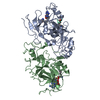
| ||||||||
|---|---|---|---|---|---|---|---|---|---|
| 1 | 
| ||||||||
| 2 | 
| ||||||||
| 3 |
| ||||||||
| Unit cell |
|
-Components
| #1: Protein | Mass: 32830.219 Da / Num. of mol.: 2 / Fragment: unp residues 982-1266 Source method: isolated from a genetically manipulated source Source: (gene. exp.) Homo sapiens (human) / Gene: EHMT1, EUHMTASE1, GLP, KIAA1876, KMT1D / Plasmid: pXC681 / Production host:  References: UniProt: Q9H9B1, Transferases; Transferring one-carbon groups; Methyltransferases, histone-lysine N-methyltransferase #2: Protein/peptide | Mass: 1286.509 Da / Num. of mol.: 2 / Fragment: unp residues 39-50 / Source method: obtained synthetically / Source: (synth.) References: UniProt: O88508, DNA (cytosine-5-)-methyltransferase #3: Chemical | ChemComp-ZN /   Mass: 65.409 Da / Num. of mol.: 8 / Source method: obtained synthetically / Formula: Zn Mass: 65.409 Da / Num. of mol.: 8 / Source method: obtained synthetically / Formula: Zn#4: Chemical |   Type: L-peptide linking / Mass: 384.411 Da / Num. of mol.: 2 / Source method: obtained synthetically / Formula: C14H20N6O5S Type: L-peptide linking / Mass: 384.411 Da / Num. of mol.: 2 / Source method: obtained synthetically / Formula: C14H20N6O5S#5: Water | ChemComp-HOH / |  Mass: 18.015 Da / Num. of mol.: 170 / Source method: isolated from a natural source / Formula: H2O Mass: 18.015 Da / Num. of mol.: 170 / Source method: isolated from a natural source / Formula: H2O |
|---|
-Experimental details
-Experiment
| Experiment | Method: X-RAY DIFFRACTION / Number of used crystals: 1 |
|---|
- Sample preparation
| Crystal | Density Matthews: 2.53 Å3/Da / Density % sol: 51.3 % |
|---|---|
| Crystal grow | Temperature: 289 K / Method: vapor diffusion, hanging drop / pH: 7.5 Details: 0.1 M Hepes, 16% polyethylene glycol 4000 and 8% isopropanol, pH 7.5, VAPOR DIFFUSION, HANGING DROP, temperature 289K |
-Data collection
| Diffraction | Mean temperature: 100 K |
|---|---|
| Diffraction source | Source: SYNCHROTRON / Site: APS  / Beamline: 24-ID-C / Wavelength: 0.97949 Å / Beamline: 24-ID-C / Wavelength: 0.97949 Å |
| Detector | Type: ADSC QUANTUM 315 / Detector: CCD / Date: Feb 7, 2010 |
| Radiation | Monochromator: Si(111) / Protocol: SINGLE WAVELENGTH / Monochromatic (M) / Laue (L): M / Scattering type: x-ray |
| Radiation wavelength | Wavelength: 0.97949 Å / Relative weight: 1 |
| Reflection | Resolution: 2.33→40.78 Å / Num. obs: 29318 / % possible obs: 98.1 % / Observed criterion σ(I): -3 / Redundancy: 8.8 % / Rmerge(I) obs: 0.072 / Net I/σ(I): 26 |
| Reflection shell | Resolution: 2.33→2.4 Å / Redundancy: 8.7 % / Rmerge(I) obs: 0.406 / Mean I/σ(I) obs: 5.6 / Num. unique all: 2825 / % possible all: 95.9 |
- Processing
| Software |
| |||||||||||||||||||||||||||||||||||||||||||||||||||||||||||||||||||||||||||||
|---|---|---|---|---|---|---|---|---|---|---|---|---|---|---|---|---|---|---|---|---|---|---|---|---|---|---|---|---|---|---|---|---|---|---|---|---|---|---|---|---|---|---|---|---|---|---|---|---|---|---|---|---|---|---|---|---|---|---|---|---|---|---|---|---|---|---|---|---|---|---|---|---|---|---|---|---|---|---|
| Refinement | Method to determine structure: MOLECULAR REPLACEMENT Starting model: PDB 3MO5 Resolution: 2.332→40.78 Å / SU ML: 0.34 / Cross valid method: THROUGHOUT / σ(F): 0.04 / Phase error: 22.5 / Stereochemistry target values: ML
| |||||||||||||||||||||||||||||||||||||||||||||||||||||||||||||||||||||||||||||
| Solvent computation | Shrinkage radii: 0.9 Å / VDW probe radii: 1.11 Å / Solvent model: FLAT BULK SOLVENT MODEL / Bsol: 31.413 Å2 / ksol: 0.354 e/Å3 | |||||||||||||||||||||||||||||||||||||||||||||||||||||||||||||||||||||||||||||
| Displacement parameters |
| |||||||||||||||||||||||||||||||||||||||||||||||||||||||||||||||||||||||||||||
| Refinement step | Cycle: LAST / Resolution: 2.332→40.78 Å
| |||||||||||||||||||||||||||||||||||||||||||||||||||||||||||||||||||||||||||||
| Refine LS restraints |
| |||||||||||||||||||||||||||||||||||||||||||||||||||||||||||||||||||||||||||||
| LS refinement shell | Refine-ID: X-RAY DIFFRACTION
|
