 Movie
Movie Controller


+ Open data

- Basic information
| Entry | Database: PDB / ID: 3siq | ||||||
|---|---|---|---|---|---|---|---|
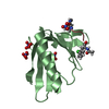
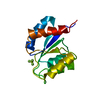
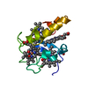
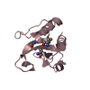
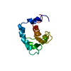
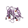
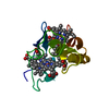
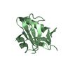
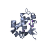
| Title | Crystal Structure of autoinhibited dIAP1-BIR1 domain | ||||||
 Components Components | Apoptosis 1 inhibitor | ||||||
 Keywords Keywords | LIGASE / dIAP1-BIR1 domain | ||||||
| Function / homology |  Function and homology information Function and homology informationSMAC, XIAP-regulated apoptotic response / DS ligand bound to FT receptor / negative regulation of compound eye retinal cell programmed cell death / antennal morphogenesis / Deactivation of the beta-catenin transactivating complex / Regulation of necroptotic cell death / Regulation of PTEN localization / sensory organ precursor cell division / Regulation of the apoptosome activity / Activation of caspases through apoptosome-mediated cleavage ...SMAC, XIAP-regulated apoptotic response / DS ligand bound to FT receptor / negative regulation of compound eye retinal cell programmed cell death / antennal morphogenesis / Deactivation of the beta-catenin transactivating complex / Regulation of necroptotic cell death / Regulation of PTEN localization / sensory organ precursor cell division / Regulation of the apoptosome activity / Activation of caspases through apoptosome-mediated cleavage / Regulation of PTEN stability and activity / spermatid nucleus differentiation / border follicle cell migration / positive regulation of Toll signaling pathway / positive regulation of border follicle cell migration / chaeta development / caspase binding / cysteine-type endopeptidase inhibitor activity involved in apoptotic process / protein neddylation / negative regulation of JNK cascade / ubiquitin conjugating enzyme binding / NEDD8 ligase activity / ubiquitin-like protein conjugating enzyme binding / ubiquitin-specific protease binding / cysteine-type endopeptidase inhibitor activity / protein autoubiquitination / protein K48-linked ubiquitination / positive regulation of protein ubiquitination / RING-type E3 ubiquitin transferase / Wnt signaling pathway / protein polyubiquitination / ubiquitin-protein transferase activity / positive regulation of canonical Wnt signaling pathway / ubiquitin protein ligase activity / spermatogenesis / regulation of cell cycle / apoptotic process / ubiquitin protein ligase binding / negative regulation of apoptotic process / perinuclear region of cytoplasm / zinc ion binding / nucleus / cytosol / cytoplasm Similarity search - Function | ||||||
| Biological species |  | ||||||
| Method |  X-RAY DIFFRACTION / SYNCHROTRON / MOLECULAR REPLACEMENT / Resolution: 2.4 Å X-RAY DIFFRACTION / SYNCHROTRON / MOLECULAR REPLACEMENT / Resolution: 2.4 Å | ||||||
 Authors Authors | Li, X. / Wang, J. / Shi, Y. | ||||||
 Citation Citation | Journal: Nat Commun / Year: 2011 Title: Structural mechanisms of DIAP1 auto-inhibition and DIAP1-mediated inhibition of drICE. Authors: Li, X. / Wang, J. / Shi, Y. | ||||||
| History |
|
- Structure visualization
| Structure viewer | Molecule: MolmilJmol/JSmol |
|---|
- Downloads & links
-Download
| PDBx/mmCIF format | 3siq.cif.gz | 132.7 KB | Display | PDBx/mmCIF format |
|---|---|---|---|---|
| PDB format | pdb3siq.ent.gz | 103.3 KB | Display | PDB format |
| PDBx/mmJSON format | 3siq.json.gz | Tree view | PDBx/mmJSON format | |
| Others |  Other downloads Other downloads |
-Validation report
| Arichive directory | https://data.pdbj.org/pub/pdb/validation_reports/si/3siqftp://data.pdbj.org/pub/pdb/validation_reports/si/3siq | HTTPS FTP |
|---|
-Related structure data
| Related structure data |  3sipC  3sirC  1sdzS C: citing same article ( S: Starting model for refinement |
|---|---|
| Similar structure data |
-Links
 PDBj
PDBj


- Assembly
| Deposited unit | 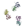
| ||||||||||||||||||||||||||||||||||||||||||||||||||||||||||||||||||
|---|---|---|---|---|---|---|---|---|---|---|---|---|---|---|---|---|---|---|---|---|---|---|---|---|---|---|---|---|---|---|---|---|---|---|---|---|---|---|---|---|---|---|---|---|---|---|---|---|---|---|---|---|---|---|---|---|---|---|---|---|---|---|---|---|---|---|---|
| 1 | 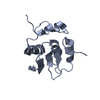
| ||||||||||||||||||||||||||||||||||||||||||||||||||||||||||||||||||
| 2 | 
| ||||||||||||||||||||||||||||||||||||||||||||||||||||||||||||||||||
| 3 | 
| ||||||||||||||||||||||||||||||||||||||||||||||||||||||||||||||||||
| 4 | 
| ||||||||||||||||||||||||||||||||||||||||||||||||||||||||||||||||||
| 5 | 
| ||||||||||||||||||||||||||||||||||||||||||||||||||||||||||||||||||
| 6 | 
| ||||||||||||||||||||||||||||||||||||||||||||||||||||||||||||||||||
| Unit cell |
| ||||||||||||||||||||||||||||||||||||||||||||||||||||||||||||||||||
| Noncrystallographic symmetry (NCS) | NCS domain:
NCS domain segments:
NCS ensembles :
|
-Components
| #1: Protein | Mass: 15996.921 Da / Num. of mol.: 6 / Fragment: BIR1 domain (UNP RESIDIES 1-136) / Mutation: C89S Source method: isolated from a genetically manipulated source Source: (gene. exp.) References: UniProt: Q24306, Ligases; Forming carbon-nitrogen bonds; Acid-amino-acid ligases (peptide synthases) #2: Chemical | ChemComp-ZN /   Mass: 65.409 Da / Num. of mol.: 6 / Source method: obtained synthetically / Formula: Zn Mass: 65.409 Da / Num. of mol.: 6 / Source method: obtained synthetically / Formula: Zn#3: Water | ChemComp-HOH / |  Mass: 18.015 Da / Num. of mol.: 4 / Source method: isolated from a natural source / Formula: H2O Mass: 18.015 Da / Num. of mol.: 4 / Source method: isolated from a natural source / Formula: H2O |
|---|
-Experimental details
-Experiment
| Experiment | Method: X-RAY DIFFRACTION / Number of used crystals: 1 |
|---|
- Sample preparation
| Crystal | Density Matthews: 2.14 Å3/Da / Density % sol: 42.43 % |
|---|---|
| Crystal grow | Temperature: 291 K / Method: vapor diffusion, hanging drop / pH: 5.4 Details: 100mM Bis-Tris pH 5.4, 0.2M (NH4)2SO4, 3%( vol/vol) CH3OH, 22% (wt/vol) Polyethylene Glycol 3350 , VAPOR DIFFUSION, HANGING DROP, temperature 291K |
-Data collection
| Diffraction | Mean temperature: 100 K | |||||||||||||||||||||||||
|---|---|---|---|---|---|---|---|---|---|---|---|---|---|---|---|---|---|---|---|---|---|---|---|---|---|---|
| Diffraction source | Source: SYNCHROTRON / Site: SPring-8  / Beamline: BL41XU / Beamline: BL41XU | |||||||||||||||||||||||||
| Detector | Type: MARMOSAIC 225 mm CCD / Detector: CCD / Date: Jul 16, 2009 | |||||||||||||||||||||||||
| Radiation | Protocol: SINGLE WAVELENGTH / Monochromatic (M) / Laue (L): M / Scattering type: x-ray | |||||||||||||||||||||||||
| Radiation wavelength | Relative weight: 1 | |||||||||||||||||||||||||
| Reflection twin |
| |||||||||||||||||||||||||
| Reflection | Resolution: 2.4→50 Å / Num. obs: 30880 / % possible obs: 99.4 % / Observed criterion σ(F): 11.5 / Observed criterion σ(I): 2.3 |
- Processing
| Software | Name: REFMAC / Version: 5.5.0109 / Classification: refinement | ||||||||||||||||||||||||||||||||||||||||||||||||||||||||||||||||||||||||||||||||||||||||||||||||||||||||||||||||||||||||||||||||||||||||||||||||||||||||||||||||||||||||||
|---|---|---|---|---|---|---|---|---|---|---|---|---|---|---|---|---|---|---|---|---|---|---|---|---|---|---|---|---|---|---|---|---|---|---|---|---|---|---|---|---|---|---|---|---|---|---|---|---|---|---|---|---|---|---|---|---|---|---|---|---|---|---|---|---|---|---|---|---|---|---|---|---|---|---|---|---|---|---|---|---|---|---|---|---|---|---|---|---|---|---|---|---|---|---|---|---|---|---|---|---|---|---|---|---|---|---|---|---|---|---|---|---|---|---|---|---|---|---|---|---|---|---|---|---|---|---|---|---|---|---|---|---|---|---|---|---|---|---|---|---|---|---|---|---|---|---|---|---|---|---|---|---|---|---|---|---|---|---|---|---|---|---|---|---|---|---|---|---|---|---|---|
| Refinement | Method to determine structure: MOLECULAR REPLACEMENT Starting model: 1SDZ Resolution: 2.4→32.98 Å / Cor.coef. Fo:Fc: 0.952 / Cor.coef. Fo:Fc free: 0.913 / SU B: 8.816 / SU ML: 0.194 / Cross valid method: THROUGHOUT / ESU R Free: 0.046 / Stereochemistry target values: MAXIMUM LIKELIHOOD / Details: HYDROGENS HAVE BEEN ADDED IN THE RIDING POSITIONS
| ||||||||||||||||||||||||||||||||||||||||||||||||||||||||||||||||||||||||||||||||||||||||||||||||||||||||||||||||||||||||||||||||||||||||||||||||||||||||||||||||||||||||||
| Solvent computation | Ion probe radii: 0.8 Å / Shrinkage radii: 0.8 Å / VDW probe radii: 1.4 Å / Solvent model: MASK | ||||||||||||||||||||||||||||||||||||||||||||||||||||||||||||||||||||||||||||||||||||||||||||||||||||||||||||||||||||||||||||||||||||||||||||||||||||||||||||||||||||||||||
| Displacement parameters | Biso mean: 29.7 Å2
| ||||||||||||||||||||||||||||||||||||||||||||||||||||||||||||||||||||||||||||||||||||||||||||||||||||||||||||||||||||||||||||||||||||||||||||||||||||||||||||||||||||||||||
| Refinement step | Cycle: LAST / Resolution: 2.4→32.98 Å
| ||||||||||||||||||||||||||||||||||||||||||||||||||||||||||||||||||||||||||||||||||||||||||||||||||||||||||||||||||||||||||||||||||||||||||||||||||||||||||||||||||||||||||
| Refine LS restraints |
| ||||||||||||||||||||||||||||||||||||||||||||||||||||||||||||||||||||||||||||||||||||||||||||||||||||||||||||||||||||||||||||||||||||||||||||||||||||||||||||||||||||||||||
| Refine LS restraints NCS | Refine-ID: X-RAY DIFFRACTION
| ||||||||||||||||||||||||||||||||||||||||||||||||||||||||||||||||||||||||||||||||||||||||||||||||||||||||||||||||||||||||||||||||||||||||||||||||||||||||||||||||||||||||||
| LS refinement shell | Resolution: 2.399→2.461 Å / Total num. of bins used: 20
|
