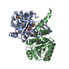
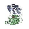
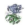
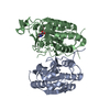
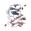
Regulation by c-FLIP / Breakdown of the nuclear lamina / Caspase activation via Dependence Receptors in the absence of ligand / CASP8 activity is inhibited / Dimerization of procaspase-8 / Apoptotic execution phase / Regulation of NF-kappa B signaling / Apoptotic cleavage of cellular proteins / Signaling by Hippo / Caspase-mediated cleavage of cytoskeletal proteins ...Regulation by c-FLIP / Breakdown of the nuclear lamina / Caspase activation via Dependence Receptors in the absence of ligand / CASP8 activity is inhibited / Dimerization of procaspase-8 / Apoptotic execution phase / Regulation of NF-kappa B signaling / Apoptotic cleavage of cellular proteins / Signaling by Hippo / Caspase-mediated cleavage of cytoskeletal proteins / Apoptosis induced DNA fragmentation / BIR domain binding / Apoptotic cleavage of cell adhesion proteins / negative regulation of Toll signaling pathway / SMAC, XIAP-regulated apoptotic response / nurse cell apoptotic process / Regulation of TNFR1 signaling / salivary gland histolysis / Regulation of necroptotic cell death / negative regulation of peptidoglycan recognition protein signaling pathway / Activation of caspases through apoptosome-mediated cleavage / compound eye retinal cell programmed cell death / spermatid differentiation / programmed cell death involved in cell development / programmed cell death / execution phase of apoptosis / neuron remodeling / response to X-ray / positive regulation of neuron apoptotic process / Hydrolases; Acting on peptide bonds (peptidases); Cysteine endopeptidases / cysteine-type endopeptidase activity / apoptotic process / ubiquitin protein ligase binding / proteolysis / nucleus / plasma membrane / cytoplasm Similarity search - Function
Rossmann fold - #1460 / Peptidase C14 family / Peptidase family C14A, His active site / Caspase family histidine active site. / Peptidase C14, caspase non-catalytic subunit p10 / Peptidase family C14A, cysteine active site / Caspase family cysteine active site. / Caspase family p10 domain profile. / Peptidase C14A, caspase catalytic domain / Caspase, interleukin-1 beta converting enzyme (ICE) homologues ...Rossmann fold - #1460 / Peptidase C14 family / Peptidase family C14A, His active site / Caspase family histidine active site. / Peptidase C14, caspase non-catalytic subunit p10 / Peptidase family C14A, cysteine active site / Caspase family cysteine active site. / Caspase family p10 domain profile. / Peptidase C14A, caspase catalytic domain / Caspase, interleukin-1 beta converting enzyme (ICE) homologues / Peptidase C14, p20 domain / Caspase family p20 domain profile. / : / Caspase domain / Caspase-like domain superfamily / Rossmann fold / 3-Layer(aba) Sandwich / Alpha Beta Similarity search - Domain/homology
Resolution: 2.68→45.97 Å / Cor.coef. Fo:Fc: 0.944 / Cor.coef. Fo:Fc free: 0.889 / SU B: 8.87 / SU ML: 0.191 / Cross valid method: THROUGHOUT / ESU R: 0.124 / ESU R Free: 0.064 / Stereochemistry target values: MAXIMUM LIKELIHOOD / Details: HYDROGENS HAVE BEEN ADDED IN THE RIDING POSITIONS
Rfactor
Num. reflection
% reflection
Selection details
Rfree
0.23517
1400
5 %
RANDOM
Rwork
0.16508
-
-
-
obs
0.1684
26529
98.58 %
-
Solvent computation
Ion probe radii: 0.8 Å / Shrinkage radii: 0.8 Å / VDW probe radii: 1.4 Å / Solvent model: MASK
Displacement parameters
Biso mean: 43.349 Å2
Baniso -1
Baniso -2
Baniso -3
1-
-14.79 Å2
0 Å2
0 Å2
2-
-
-14.79 Å2
0 Å2
3-
-
-
29.58 Å2
Refinement step
Cycle: LAST / Resolution: 2.68→45.97 Å
Protein
Nucleic acid
Ligand
Solvent
Total
Num. atoms
5763
0
0
3
5766
Refine LS restraints
Refine-ID
Type
Dev ideal
Dev ideal target
Number
X-RAY DIFFRACTION
r_bond_refined_d
0.003
0.022
5873
X-RAY DIFFRACTION
r_bond_other_d
X-RAY DIFFRACTION
r_angle_refined_deg
0.714
1.939
7925
X-RAY DIFFRACTION
r_angle_other_deg
X-RAY DIFFRACTION
r_dihedral_angle_1_deg
12.709
5
695
X-RAY DIFFRACTION
r_dihedral_angle_2_deg
40.088
23.164
275
X-RAY DIFFRACTION
r_dihedral_angle_3_deg
25.652
15
1013
X-RAY DIFFRACTION
r_dihedral_angle_4_deg
24.667
15
42
X-RAY DIFFRACTION
r_chiral_restr
0.056
0.2
908
X-RAY DIFFRACTION
r_gen_planes_refined
0.015
0.02
4340
X-RAY DIFFRACTION
r_gen_planes_other
X-RAY DIFFRACTION
r_nbd_refined
X-RAY DIFFRACTION
r_nbd_other
X-RAY DIFFRACTION
r_nbtor_refined
X-RAY DIFFRACTION
r_nbtor_other
X-RAY DIFFRACTION
r_xyhbond_nbd_refined
X-RAY DIFFRACTION
r_xyhbond_nbd_other
X-RAY DIFFRACTION
r_metal_ion_refined
X-RAY DIFFRACTION
r_metal_ion_other
X-RAY DIFFRACTION
r_symmetry_vdw_refined
X-RAY DIFFRACTION
r_symmetry_vdw_other
X-RAY DIFFRACTION
r_symmetry_hbond_refined
X-RAY DIFFRACTION
r_symmetry_hbond_other
X-RAY DIFFRACTION
r_symmetry_metal_ion_refined
X-RAY DIFFRACTION
r_symmetry_metal_ion_other
X-RAY DIFFRACTION
r_mcbond_it
1.263
1.5
3559
X-RAY DIFFRACTION
r_mcbond_other
X-RAY DIFFRACTION
r_mcangle_it
2.367
2
5738
X-RAY DIFFRACTION
r_scbond_it
3.666
3
2314
X-RAY DIFFRACTION
r_scangle_it
5.683
4.5
2187
X-RAY DIFFRACTION
r_rigid_bond_restr
X-RAY DIFFRACTION
r_sphericity_free
X-RAY DIFFRACTION
r_sphericity_bonded
LS refinement shell
Resolution: 2.68→2.749 Å / Total num. of bins used: 20
Rfactor
Num. reflection
% reflection
Rfree
0
0
-
Rwork
0.218
1863
-
obs
-
-
89.91 %
+
About Yorodumi
-
News
-
Feb 9, 2022. New format data for meta-information of EMDB entries
New format data for meta-information of EMDB entries
Version 3 of the EMDB header file is now the official format.
The previous official version 1.9 will be removed from the archive.
In the structure databanks used in Yorodumi, some data are registered as the other names, "COVID-19 virus" and "2019-nCoV". Here are the details of the virus and the list of structure data.
Jan 31, 2019. EMDB accession codes are about to change! (news from PDBe EMDB page)
EMDB accession codes are about to change! (news from PDBe EMDB page)
The allocation of 4 digits for EMDB accession codes will soon come to an end. Whilst these codes will remain in use, new EMDB accession codes will include an additional digit and will expand incrementally as the available range of codes is exhausted. The current 4-digit format prefixed with “EMD-” (i.e. EMD-XXXX) will advance to a 5-digit format (i.e. EMD-XXXXX), and so on. It is currently estimated that the 4-digit codes will be depleted around Spring 2019, at which point the 5-digit format will come into force.
The EM Navigator/Yorodumi systems omit the EMD- prefix.
Related info.:Q: What is EMD? / ID/Accession-code notation in Yorodumi/EM Navigator
Yorodumi is a browser for structure data from EMDB, PDB, SASBDB, etc.
This page is also the successor to EM Navigator detail page, and also detail information page/front-end page for Omokage search.
The word "yorodu" (or yorozu) is an old Japanese word meaning "ten thousand". "mi" (miru) is to see.
Related info.:EMDB / PDB / SASBDB / Comparison of 3 databanks / Yorodumi Search / Aug 31, 2016. New EM Navigator & Yorodumi / Yorodumi Papers / Jmol/JSmol / Function and homology information / Changes in new EM Navigator and Yorodumi
 Movie
Movie Controller


 Open data
 Basic information
 Components
 Keywords
 Function and homology information
 X-RAY DIFFRACTION /
  Authors
 Citation
 Structure visualization
 Downloads & links
 Other downloads


 PDBj
PDBj

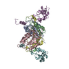
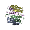
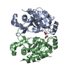
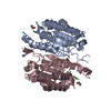
 Assembly


 Mass: 18.015 Da / Num. of mol.: 3 / Source method: isolated from a natural source / Formula: H2O
 Sample preparation
 / Beamline: BL38B1
 Processing