 Movie
Movie Controller


+ Open data

- Basic information
| Entry | Database: PDB / ID: 5hdk | ||||||
|---|---|---|---|---|---|---|---|
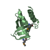
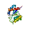
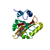
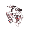
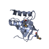
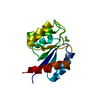
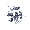
| Title | Crystal structure of heat shock factor 2-DBD | ||||||
 Components Components | Heat shock factor protein 2 | ||||||
 Keywords Keywords | TRANSCRIPTION / HSF1-DBD | ||||||
| Function / homology |  Function and homology information Function and homology informationRNA polymerase II intronic transcription regulatory region sequence-specific DNA binding / RNA polymerase II transcription regulatory region sequence-specific DNA binding / sequence-specific double-stranded DNA binding / DNA-binding transcription activator activity, RNA polymerase II-specific / spermatogenesis / DNA-binding transcription factor activity, RNA polymerase II-specific / RNA polymerase II cis-regulatory region sequence-specific DNA binding / chromatin / positive regulation of transcription by RNA polymerase II / nucleoplasm ...RNA polymerase II intronic transcription regulatory region sequence-specific DNA binding / RNA polymerase II transcription regulatory region sequence-specific DNA binding / sequence-specific double-stranded DNA binding / DNA-binding transcription activator activity, RNA polymerase II-specific / spermatogenesis / DNA-binding transcription factor activity, RNA polymerase II-specific / RNA polymerase II cis-regulatory region sequence-specific DNA binding / chromatin / positive regulation of transcription by RNA polymerase II / nucleoplasm / identical protein binding / cytoplasm Similarity search - Function | ||||||
| Biological species |  Homo sapiens (human) Homo sapiens (human) | ||||||
| Method |  X-RAY DIFFRACTION / SYNCHROTRON / MOLECULAR REPLACEMENT / Resolution: 1.32 Å X-RAY DIFFRACTION / SYNCHROTRON / MOLECULAR REPLACEMENT / Resolution: 1.32 Å | ||||||
 Authors Authors | Feng, H. / Liu, W. / Wang, D.C. | ||||||
 Citation Citation | Journal: To Be Published Title: HSF1-DBD crystal structure Authors: Feng, H. / Liu, W. / Wang, D.C. | ||||||
| History |
|
- Structure visualization
| Structure viewer | Molecule: MolmilJmol/JSmol |
|---|
- Downloads & links
-Download
| PDBx/mmCIF format | 5hdk.cif.gz | 202.4 KB | Display | PDBx/mmCIF format |
|---|---|---|---|---|
| PDB format | pdb5hdk.ent.gz | 159.7 KB | Display | PDB format |
| PDBx/mmJSON format | 5hdk.json.gz | Tree view | PDBx/mmJSON format | |
| Others |  Other downloads Other downloads |
-Validation report
| Arichive directory | https://data.pdbj.org/pub/pdb/validation_reports/hd/5hdkftp://data.pdbj.org/pub/pdb/validation_reports/hd/5hdk | HTTPS FTP |
|---|
-Related structure data
| Related structure data |  5hdgSC  5hdnC S: Starting model for refinement C: citing same article ( |
|---|---|
| Similar structure data |


-Links
 PDBj
PDBj- Assembly
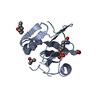
| Deposited unit | 
| ||||||||
|---|---|---|---|---|---|---|---|---|---|
| 1 | 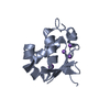
| ||||||||
| 2 | 
| ||||||||
| 3 | 
| ||||||||
| 4 | 
| ||||||||
| Unit cell |
|
-Components
| #1: Protein | Mass: 13405.185 Da / Num. of mol.: 4 / Fragment: UNP residues 7-112 Source method: isolated from a genetically manipulated source Source: (gene. exp.) Homo sapiens (human) / Gene: HSF2, HSTF2 / Production host:  #2: Chemical | ChemComp-K /   Mass: 39.098 Da / Num. of mol.: 6 / Source method: obtained synthetically / Formula: K Mass: 39.098 Da / Num. of mol.: 6 / Source method: obtained synthetically / Formula: K#3: Chemical | ChemComp-NA /   Mass: 22.990 Da / Num. of mol.: 9 / Source method: obtained synthetically / Formula: Na Mass: 22.990 Da / Num. of mol.: 9 / Source method: obtained synthetically / Formula: Na#4: Chemical |   Mass: 35.453 Da / Num. of mol.: 3 / Source method: obtained synthetically / Formula: Cl Mass: 35.453 Da / Num. of mol.: 3 / Source method: obtained synthetically / Formula: Cl#5: Water | ChemComp-HOH / |  Mass: 18.015 Da / Num. of mol.: 543 / Source method: isolated from a natural source / Formula: H2O Mass: 18.015 Da / Num. of mol.: 543 / Source method: isolated from a natural source / Formula: H2O |
|---|
-Experimental details
-Experiment
| Experiment | Method: X-RAY DIFFRACTION |
|---|
- Sample preparation
| Crystal | Density Matthews: 1.92 Å3/Da / Density % sol: 35.94 % |
|---|---|
| Crystal grow | Temperature: 293 K / Method: vapor diffusion, hanging drop / pH: 8.4 Details: 0.1 M Tris, pH 8.4 and 1.25 M potassium sodium tartrate |
-Data collection
| Diffraction | Mean temperature: 100 K |
|---|---|
| Diffraction source | Source: SYNCHROTRON / Site: SSRF  / Beamline: BL19U1 / Wavelength: 0.97845 Å / Beamline: BL19U1 / Wavelength: 0.97845 Å |
| Detector | Type: DECTRIS PILATUS3 6M / Detector: PIXEL / Date: May 3, 2015 |
| Radiation | Protocol: SINGLE WAVELENGTH / Monochromatic (M) / Laue (L): M / Scattering type: x-ray |
| Radiation wavelength | Wavelength: 0.97845 Å / Relative weight: 1 |
| Reflection | Resolution: 1.32→38.32 Å / Num. obs: 95303 / % possible obs: 99.8 % / Redundancy: 12 % / Rmerge(I) obs: 0.066 / Rsym value: 0.066 / Net I/σ(I): 3.6 |
| Reflection shell | Resolution: 1.32→1.39 Å / Redundancy: 11.2 % / Rmerge(I) obs: 0.657 / Mean I/σ(I) obs: 3 / % possible all: 99.8 |
- Processing
| Software |
| |||||||||||||||||||||||||||||||||||||||||||||||||||||||||||||||||||||||||||||||||||||||||||||||||||||||||
|---|---|---|---|---|---|---|---|---|---|---|---|---|---|---|---|---|---|---|---|---|---|---|---|---|---|---|---|---|---|---|---|---|---|---|---|---|---|---|---|---|---|---|---|---|---|---|---|---|---|---|---|---|---|---|---|---|---|---|---|---|---|---|---|---|---|---|---|---|---|---|---|---|---|---|---|---|---|---|---|---|---|---|---|---|---|---|---|---|---|---|---|---|---|---|---|---|---|---|---|---|---|---|---|---|---|---|
| Refinement | Method to determine structure: MOLECULAR REPLACEMENT Starting model: 5HDG Resolution: 1.32→38.319 Å / SU ML: 0.1 / Cross valid method: NONE / σ(F): 0 / Phase error: 16.53 / Stereochemistry target values: ML
| |||||||||||||||||||||||||||||||||||||||||||||||||||||||||||||||||||||||||||||||||||||||||||||||||||||||||
| Solvent computation | Shrinkage radii: 0.9 Å / VDW probe radii: 1.11 Å / Solvent model: FLAT BULK SOLVENT MODEL | |||||||||||||||||||||||||||||||||||||||||||||||||||||||||||||||||||||||||||||||||||||||||||||||||||||||||
| Refinement step | Cycle: LAST / Resolution: 1.32→38.319 Å
| |||||||||||||||||||||||||||||||||||||||||||||||||||||||||||||||||||||||||||||||||||||||||||||||||||||||||
| Refine LS restraints |
| |||||||||||||||||||||||||||||||||||||||||||||||||||||||||||||||||||||||||||||||||||||||||||||||||||||||||
| LS refinement shell |
|
