 Movie
Movie Controller


+ Open data

- Basic information
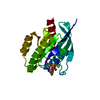
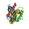
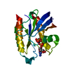
| Entry | Database: PDB / ID: 3sea | ||||||
|---|---|---|---|---|---|---|---|
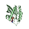
| Title | Structure of Rheb-Y35A mutant in GDP- and GMPPNP-bound forms | ||||||
 Components Components | GTP-binding protein Rheb | ||||||
 Keywords Keywords | HYDROLASE / globular | ||||||
| Function / homology |  Function and homology information Function and homology informationregulation of type B pancreatic cell development / MTOR signalling / Energy dependent regulation of mTOR by LKB1-AMPK / Amino acids regulate mTORC1 / negative regulation of cold-induced thermogenesis / small GTPase-mediated signal transduction / Macroautophagy / oligodendrocyte differentiation / positive regulation of oligodendrocyte differentiation / mTORC1-mediated signalling ...regulation of type B pancreatic cell development / MTOR signalling / Energy dependent regulation of mTOR by LKB1-AMPK / Amino acids regulate mTORC1 / negative regulation of cold-induced thermogenesis / small GTPase-mediated signal transduction / Macroautophagy / oligodendrocyte differentiation / positive regulation of oligodendrocyte differentiation / mTORC1-mediated signalling / regulation of macroautophagy / positive regulation of TOR signaling / protein kinase activator activity / endomembrane system / positive regulation of TORC1 signaling / cellular response to nutrient levels / Regulation of PTEN gene transcription / protein serine/threonine kinase activator activity / TP53 Regulates Metabolic Genes / spliceosomal complex / response to virus / GDP binding / Hydrolases; Acting on acid anhydrides; Acting on GTP to facilitate cellular and subcellular movement / regulation of cell cycle / postsynaptic density / Golgi membrane / lysosomal membrane / GTPase activity / endoplasmic reticulum membrane / protein kinase binding / GTP binding / magnesium ion binding / signal transduction / extracellular exosome / membrane / plasma membrane / cytosol Similarity search - Function | ||||||
| Biological species |  Homo sapiens (human) Homo sapiens (human) | ||||||
| Method |  X-RAY DIFFRACTION / MOLECULAR REPLACEMENT / Resolution: 2 Å X-RAY DIFFRACTION / MOLECULAR REPLACEMENT / Resolution: 2 Å | ||||||
 Authors Authors | Mazhab-Jafari, M.T. / Marshall, C.B. / Ishiyama, N. / Vuk, S. / Ikura, M. | ||||||
 Citation Citation | Journal: Structure / Year: 2012 Title: An Autoinhibited Noncanonical Mechanism of GTP Hydrolysis by Rheb Maintains mTORC1 Homeostasis. Authors: Mazhab-Jafari, M.T. / Marshall, C.B. / Ishiyama, N. / Ho, J. / Di Palma, V. / Stambolic, V. / Ikura, M. | ||||||
| History |
|
- Structure visualization
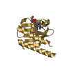
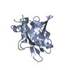
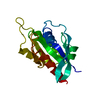
| Structure viewer | Molecule: MolmilJmol/JSmol |
|---|
- Downloads & links
-Download
| PDBx/mmCIF format | 3sea.cif.gz | 91.1 KB | Display | PDBx/mmCIF format |
|---|---|---|---|---|
| PDB format | pdb3sea.ent.gz | 68.1 KB | Display | PDB format |
| PDBx/mmJSON format | 3sea.json.gz | Tree view | PDBx/mmJSON format | |
| Others |  Other downloads Other downloads |
-Validation report
| Arichive directory | https://data.pdbj.org/pub/pdb/validation_reports/se/3seaftp://data.pdbj.org/pub/pdb/validation_reports/se/3sea | HTTPS FTP |
|---|
-Related structure data
| Related structure data | 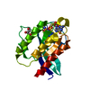 1xtrS S: Starting model for refinement |
|---|---|
| Similar structure data |


-Links
 PDBj
PDBj


- Assembly
| Deposited unit | 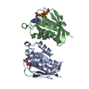
| ||||||||
|---|---|---|---|---|---|---|---|---|---|
| 1 | 
| ||||||||
| 2 | 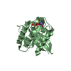
| ||||||||
| 3 | 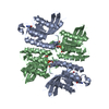
| ||||||||
| Unit cell |
|
-Components
-Protein , 1 types, 2 molecules AB
| #1: Protein | Mass: 18757.406 Da / Num. of mol.: 2 / Fragment: G-domain / Mutation: Y35A Source method: isolated from a genetically manipulated source Source: (gene. exp.) Homo sapiens (human) / Gene: RHEB, RHEB2 / Production host:  |
|---|
-Non-polymers , 5 types, 257 molecules 


| #2: Chemical |  Mass: 24.305 Da / Num. of mol.: 2 / Source method: obtained synthetically / Formula: Mg Mass: 24.305 Da / Num. of mol.: 2 / Source method: obtained synthetically / Formula: Mg#3: Chemical | ChemComp-GDP / |  Type: RNA linking / Mass: 443.201 Da / Num. of mol.: 1 / Source method: obtained synthetically / Formula: C10H15N5O11P2 / Comment: GDP, energy-carrying molecule*YM Type: RNA linking / Mass: 443.201 Da / Num. of mol.: 1 / Source method: obtained synthetically / Formula: C10H15N5O11P2 / Comment: GDP, energy-carrying molecule*YM#4: Chemical | ChemComp-GNP / |  Mass: 522.196 Da / Num. of mol.: 1 / Source method: obtained synthetically / Formula: C10H17N6O13P3 Mass: 522.196 Da / Num. of mol.: 1 / Source method: obtained synthetically / Formula: C10H17N6O13P3Comment: GppNHp, GMPPNP, energy-carrying molecule analogue*YM #5: Chemical | ChemComp-ACT / |  Mass: 59.044 Da / Num. of mol.: 1 / Source method: obtained synthetically / Formula: C2H3O2 Mass: 59.044 Da / Num. of mol.: 1 / Source method: obtained synthetically / Formula: C2H3O2#6: Water | ChemComp-HOH / | Mass: 18.015 Da / Num. of mol.: 252 / Source method: isolated from a natural source / Formula: H2O |
|---|
-Experimental details
-Experiment
| Experiment | Method: X-RAY DIFFRACTION / Number of used crystals: 1 |
|---|
- Sample preparation
| Crystal | Density Matthews: 2.11 Å3/Da / Density % sol: 41.73 % |
|---|---|
| Crystal grow | Temperature: 293 K / Method: vapor diffusion, hanging drop / pH: 8.5 Details: 100 mM Tris hydrochloride, 200 mM Sodium acetate trihydrate, 30% w/v Polyethylene glycol 4,000, pH 8.5, VAPOR DIFFUSION, HANGING DROP, temperature 293K |
-Data collection
| Diffraction | Mean temperature: 100 K |
|---|---|
| Diffraction source | Source: ROTATING ANODE / Type: RIGAKU RUH3R / Wavelength: 1.54 Å |
| Radiation | Protocol: SINGLE WAVELENGTH / Monochromatic (M) / Laue (L): M / Scattering type: x-ray |
| Radiation wavelength | Wavelength: 1.54 Å / Relative weight: 1 |
| Reflection | Resolution: 2→50 Å / Num. obs: 21742 / % possible obs: 99.8 % / Observed criterion σ(F): 0 / Observed criterion σ(I): 3 / Redundancy: 7 % / Rmerge(I) obs: 0.093 / Rsym value: 0.093 / Net I/σ(I): 20.711 |
| Reflection shell | Resolution: 2→2.03 Å / Redundancy: 6.6 % / Rmerge(I) obs: 0.412 / Mean I/σ(I) obs: 4.597 / Rsym value: 0.412 / % possible all: 100 |
- Processing
| Software |
| |||||||||||||||||||||||||||||||||||||||||||||||||||||||||||||||||||||||||||||
|---|---|---|---|---|---|---|---|---|---|---|---|---|---|---|---|---|---|---|---|---|---|---|---|---|---|---|---|---|---|---|---|---|---|---|---|---|---|---|---|---|---|---|---|---|---|---|---|---|---|---|---|---|---|---|---|---|---|---|---|---|---|---|---|---|---|---|---|---|---|---|---|---|---|---|---|---|---|---|
| Refinement | Method to determine structure: MOLECULAR REPLACEMENT Starting model: PDB entry 1XTR Resolution: 2→26.919 Å / SU ML: 0.25 / σ(F): 0 / Phase error: 19.71 / Stereochemistry target values: ML
| |||||||||||||||||||||||||||||||||||||||||||||||||||||||||||||||||||||||||||||
| Solvent computation | Shrinkage radii: 0.9 Å / VDW probe radii: 1.11 Å / Solvent model: FLAT BULK SOLVENT MODEL / Bsol: 56.249 Å2 / ksol: 0.383 e/Å3 | |||||||||||||||||||||||||||||||||||||||||||||||||||||||||||||||||||||||||||||
| Displacement parameters |
| |||||||||||||||||||||||||||||||||||||||||||||||||||||||||||||||||||||||||||||
| Refinement step | Cycle: LAST / Resolution: 2→26.919 Å
| |||||||||||||||||||||||||||||||||||||||||||||||||||||||||||||||||||||||||||||
| Refine LS restraints |
| |||||||||||||||||||||||||||||||||||||||||||||||||||||||||||||||||||||||||||||
| LS refinement shell |
|
