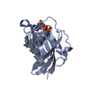
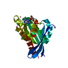
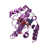
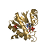
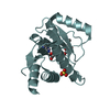
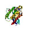
Mass: 15983.139 Da / Num. of mol.: 1 / Fragment: PTB domain, UNP residues 1263-1409 Source method: isolated from a genetically manipulated source Source: (gene. exp.) Homo sapiens (human) / Gene: TENC1 / Production host: Escherichia coli (E. coli) References: UniProt: Q63HR2, Hydrolases; Acting on ester bonds; Phosphoric-monoester hydrolases
#2: Protein/peptide
RhoGTPase-activatingprotein7 / Deleted in liver cancer 1 protein / DLC-1 / HP protein / Rho-type GTPase-activating protein 7 / ...Deleted in liver cancer 1 protein / DLC-1 / HP protein / Rho-type GTPase-activating protein 7 / START domain-containing protein 12 / StARD12 / StAR-related lipid transfer protein 12
Mass: 1557.724 Da / Num. of mol.: 1 / Fragment: UNP residues 811-824 Source method: isolated from a genetically manipulated source Source: (gene. exp.) Homo sapiens (human) / Gene: DLC1 / Production host: Escherichia coli (E. coli) / References: UniProt: Q96QB1
-
Experimental details
-
Experiment
Experiment
Method: SOLUTION NMR
NMR experiment
Conditions-ID
Experiment-ID
Solution-ID
Type
1
1
1
2D 1H-15N HSQC
1
2
2
2D 1H-15N HSQC
1
3
1
2D 1H-13C HSQC
1
4
2
2D 1H-13C HSQC
1
5
1
3D HNCO
1
6
1
3D HN(CA)CB
1
7
1
3DCBCA(CO)NH
1
8
3
3D (H)CCH-TOCSY
1
9
1
3D 1H-15N NOESY
1
10
3
3D 1H-13C NOESY
1
11
2
3D HNCO
1
12
2
3DCBCA(CO)NH
1
13
2
3D HN(CA)CB
1
14
2
3D 1H-15N NOESY
1
15
4
3D 1H-13C NOESY
1
16
4
3D (H)CCH-COSY
1
17
3
3D13C/15N FILTERED-13C EDITED NOESY
1
18
4
3D13C/15N FILTERED-13C EDITED NOESY
-
Sample preparation
Details
Solution-ID
Contents
Solvent system
1
0.6-0.8 mM [U-100% 13C; U-100% 15N] Tensin2-1, 1.2-1.4 mM DLC1-2, 100 mM potassium phosphate-3, 100 mM potassium chloride-4, 1 mM EDTA-5, 90% H2O/10% D2O
90% H2O/10% D2O
2
1.2-1.4 mM Tensin2-6, 0.6-0.8 mM [U-100% 13C; U-100% 15N] DLC1-7, 100 mM potassium phosphate-8, 100 mM potassium chloride-9, 1 mM EDTA-10, 90% H2O/10% D2O
90% H2O/10% D2O
3
0.6-0.8 mM [U-100% 13C] Tensin2-11, 1.2-1.4 mM DLC1-12, 100 mM potassium phosphate-13, 100 mM potassium chloride-14, 1 mM EDTA-15, 100% D2O
100% D2O
4
1.2-1.4 mM Tensin2-16, 0.6-0.8 mM [U-100% 13C] DLC1-17, 100 mM potassium phosphate-18, 100 mM potassium chloride-19, 1 mM EDTA-20, 100% D2O
Conformer selection criteria: structures with the least restraint violations Conformers calculated total number: 200 / Conformers submitted total number: 10 / Representative conformer: 1
+
About Yorodumi
-
News
-
Feb 9, 2022. New format data for meta-information of EMDB entries
New format data for meta-information of EMDB entries
Version 3 of the EMDB header file is now the official format.
The previous official version 1.9 will be removed from the archive.
In the structure databanks used in Yorodumi, some data are registered as the other names, "COVID-19 virus" and "2019-nCoV". Here are the details of the virus and the list of structure data.
Jan 31, 2019. EMDB accession codes are about to change! (news from PDBe EMDB page)
EMDB accession codes are about to change! (news from PDBe EMDB page)
The allocation of 4 digits for EMDB accession codes will soon come to an end. Whilst these codes will remain in use, new EMDB accession codes will include an additional digit and will expand incrementally as the available range of codes is exhausted. The current 4-digit format prefixed with “EMD-” (i.e. EMD-XXXX) will advance to a 5-digit format (i.e. EMD-XXXXX), and so on. It is currently estimated that the 4-digit codes will be depleted around Spring 2019, at which point the 5-digit format will come into force.
The EM Navigator/Yorodumi systems omit the EMD- prefix.
Related info.:Q: What is EMD? / ID/Accession-code notation in Yorodumi/EM Navigator
Yorodumi is a browser for structure data from EMDB, PDB, SASBDB, etc.
This page is also the successor to EM Navigator detail page, and also detail information page/front-end page for Omokage search.
The word "yorodu" (or yorozu) is an old Japanese word meaning "ten thousand". "mi" (miru) is to see.
Related info.:EMDB / PDB / SASBDB / Comparison of 3 databanks / Yorodumi Search / Aug 31, 2016. New EM Navigator & Yorodumi / Yorodumi Papers / Jmol/JSmol / Function and homology information / Changes in new EM Navigator and Yorodumi
 Movie
Movie Controller


 Open data
 Basic information
 Components
 Keywords
 Function and homology information
 Homo sapiens (human)
 Authors
 Citation
 Structure visualization
 Downloads & links
 Other downloads
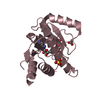


 PDBj
PDBj


 Assembly
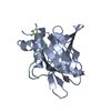

 HSQC
HSQC Sample preparation
 Processing