 Movie
Movie Controller


+ Open data

- Basic information
| Entry | Database: PDB / ID: 3se3 | ||||||
|---|---|---|---|---|---|---|---|
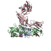
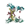
| Title | human IFNa2-IFNAR ternary complex | ||||||
 Components Components |
| ||||||
 Keywords Keywords | IMMUNE SYSTEM RECEPTOR / Type I interferon signaling complex / extracellular space | ||||||
| Function / homology |  Function and homology information Function and homology informationtype I interferon receptor activity / type I interferon binding / type I interferon receptor binding / B cell activation involved in immune response / JAK pathway signal transduction adaptor activity / response to interferon-beta / negative regulation of interleukin-5 production / negative regulation of interleukin-13 production / negative regulation of T cell differentiation / natural killer cell activation involved in immune response ...type I interferon receptor activity / type I interferon binding / type I interferon receptor binding / B cell activation involved in immune response / JAK pathway signal transduction adaptor activity / response to interferon-beta / negative regulation of interleukin-5 production / negative regulation of interleukin-13 production / negative regulation of T cell differentiation / natural killer cell activation involved in immune response / positive regulation of cellular respiration / cellular response to interferon-alpha / response to interferon-alpha / negative regulation of T-helper 2 cell cytokine production / T cell activation involved in immune response / host-mediated suppression of symbiont invasion / cytokine receptor binding / cell surface receptor signaling pathway via STAT / TRAF6 mediated IRF7 activation / type I interferon-mediated signaling pathway / response to exogenous dsRNA / cytokine binding / humoral immune response / Regulation of IFNA/IFNB signaling / cell surface receptor signaling pathway via JAK-STAT / cellular response to interferon-beta / cytokine activity / cellular response to virus / Evasion by RSV of host interferon responses / response to virus / Interferon alpha/beta signaling / late endosome / cell-cell signaling / Factors involved in megakaryocyte development and platelet production / extracellular matrix / response to lipopolysaccharide / defense response to virus / Potential therapeutics for SARS / adaptive immune response / cell surface receptor signaling pathway / lysosome / signaling receptor complex / inflammatory response / negative regulation of gene expression / negative regulation of DNA-templated transcription / apoptotic process / protein kinase binding / SARS-CoV-2 activates/modulates innate and adaptive immune responses / : / extracellular region / plasma membrane Similarity search - Function | ||||||
| Biological species |  Homo sapiens (human) Homo sapiens (human) | ||||||
| Method |  X-RAY DIFFRACTION / SYNCHROTRON / MOLECULAR REPLACEMENT / Resolution: 4.0001 Å X-RAY DIFFRACTION / SYNCHROTRON / MOLECULAR REPLACEMENT / Resolution: 4.0001 Å | ||||||
 Authors Authors | Thomas, C. / Garcia, K.C. | ||||||
 Citation Citation | Journal: Cell(Cambridge,Mass.) / Year: 2011 Title: Structural linkage between ligand discrimination and receptor activation by type I interferons. Authors: Thomas, C. / Moraga, I. / Levin, D. / Krutzik, P.O. / Podoplelova, Y. / Trejo, A. / Lee, C. / Yarden, G. / Vleck, S.E. / Glenn, J.S. / Nolan, G.P. / Piehler, J. / Schreiber, G. / Garcia, K.C. | ||||||
| History |
|
- Structure visualization
| Structure viewer | Molecule: MolmilJmol/JSmol |
|---|
- Downloads & links
-Download
| PDBx/mmCIF format | 3se3.cif.gz | 252.2 KB | Display | PDBx/mmCIF format |
|---|---|---|---|---|
| PDB format | pdb3se3.ent.gz | 200.6 KB | Display | PDB format |
| PDBx/mmJSON format | 3se3.json.gz | Tree view | PDBx/mmJSON format | |
| Others |  Other downloads Other downloads |
-Validation report
| Arichive directory | https://data.pdbj.org/pub/pdb/validation_reports/se/3se3ftp://data.pdbj.org/pub/pdb/validation_reports/se/3se3 | HTTPS FTP |
|---|
-Related structure data
| Related structure data |  3s8wC 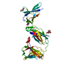 3s98C  3s9dSC 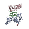 3se4SC C: citing same article ( S: Starting model for refinement |
|---|---|
| Similar structure data |


-Links
 PDBj
PDBj


- Assembly
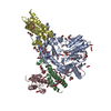
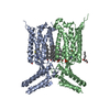
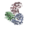
| Deposited unit | 
| ||||||||
|---|---|---|---|---|---|---|---|---|---|
| 1 |
| ||||||||
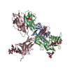
| Unit cell |
|
-Components
| #1: Protein | Mass: 47629.965 Da / Num. of mol.: 1 / Fragment: IFNa2(YNS) (UNP Residues 28-436) Source method: isolated from a genetically manipulated source Source: (gene. exp.) Homo sapiens (human) / Gene: IFNAR1, IFNAR / Production host:  Trichoplusia ni (cabbage looper) / References: UniProt: P17181 Trichoplusia ni (cabbage looper) / References: UniProt: P17181 |
|---|---|
| #2: Protein | Mass: 19392.270 Da / Num. of mol.: 1 / Mutation: H58Y, E59N, Q62S Source method: isolated from a genetically manipulated source Source: (gene. exp.) Homo sapiens (human) / Production host:  |
| #3: Protein | Mass: 22894.078 Da / Num. of mol.: 1 / Fragment: UNP Residues 34-232 Source method: isolated from a genetically manipulated source Source: (gene. exp.) Homo sapiens (human) / Gene: IFNAR2, IFNABR, IFNARB / Production host: Trichoplusia ni (cabbage looper) / References: UniProt: P48551 |
| #4: Sugar | ChemComp-NAG /   Type: D-saccharide, beta linking / Mass: 221.208 Da / Num. of mol.: 1 Type: D-saccharide, beta linking / Mass: 221.208 Da / Num. of mol.: 1Source method: isolated from a genetically manipulated source Formula: C8H15NO6 |
| Has protein modification | Y |
-Experimental details
-Experiment
| Experiment | Method: X-RAY DIFFRACTION / Number of used crystals: 1 |
|---|
- Sample preparation
| Crystal | Density Matthews: 2.81 Å3/Da / Density % sol: 56.22 % |
|---|---|
| Crystal grow | Temperature: 293 K / pH: 6.5 Details: 15% (w/v) PEG 3350, 100 mM Na malonate, pH 6.5, VAPOR DIFFUSION, HANGING DROP, temperature 293K |
-Data collection
| Diffraction | Mean temperature: 100 K |
|---|---|
| Diffraction source | Source: SYNCHROTRON / Site: ALS  / Beamline: 8.2.1 / Wavelength: 1 / Beamline: 8.2.1 / Wavelength: 1 |
| Detector | Type: ADSC QUANTUM 315r / Detector: CCD / Date: Jul 3, 2008 |
| Radiation | Protocol: SINGLE WAVELENGTH / Monochromatic (M) / Laue (L): M / Scattering type: x-ray |
| Radiation wavelength | Wavelength: 1 Å / Relative weight: 1 |
| Reflection | Resolution: 4→46.8 Å / Num. obs: 9537 / % possible obs: 99.8 % / Redundancy: 11 % / Biso Wilson estimate: 137 Å2 / Rsym value: 0.172 / Net I/σ(I): 11.7 |
| Reflection shell | Resolution: 4→4.1 Å / Redundancy: 11.5 % / Mean I/σ(I) obs: 2.6 / Rsym value: 1.03 / % possible all: 100 |
- Processing
| Software |
| ||||||||||||||||||||||||||||||||||||||||||||||||||||||||||||||||||||||||||||||||||||||||||||||||||||||||||||||||||||||||||||||||||||||||||||||||||||||||||||||||||||||||||||||||||||||||||||||||||||||||||||||||||||||||||||||||||||||||||||||||||||||||||
|---|---|---|---|---|---|---|---|---|---|---|---|---|---|---|---|---|---|---|---|---|---|---|---|---|---|---|---|---|---|---|---|---|---|---|---|---|---|---|---|---|---|---|---|---|---|---|---|---|---|---|---|---|---|---|---|---|---|---|---|---|---|---|---|---|---|---|---|---|---|---|---|---|---|---|---|---|---|---|---|---|---|---|---|---|---|---|---|---|---|---|---|---|---|---|---|---|---|---|---|---|---|---|---|---|---|---|---|---|---|---|---|---|---|---|---|---|---|---|---|---|---|---|---|---|---|---|---|---|---|---|---|---|---|---|---|---|---|---|---|---|---|---|---|---|---|---|---|---|---|---|---|---|---|---|---|---|---|---|---|---|---|---|---|---|---|---|---|---|---|---|---|---|---|---|---|---|---|---|---|---|---|---|---|---|---|---|---|---|---|---|---|---|---|---|---|---|---|---|---|---|---|---|---|---|---|---|---|---|---|---|---|---|---|---|---|---|---|---|---|---|---|---|---|---|---|---|---|---|---|---|---|---|---|---|---|---|---|---|---|---|---|---|---|---|---|---|---|---|---|---|---|
| Refinement | Method to determine structure: MOLECULAR REPLACEMENT Starting model: PDB ENTRY 3SE4, 3S9D Resolution: 4.0001→46.78 Å / σ(F): 2.01 / Stereochemistry target values: ML
| ||||||||||||||||||||||||||||||||||||||||||||||||||||||||||||||||||||||||||||||||||||||||||||||||||||||||||||||||||||||||||||||||||||||||||||||||||||||||||||||||||||||||||||||||||||||||||||||||||||||||||||||||||||||||||||||||||||||||||||||||||||||||||
| Solvent computation | Shrinkage radii: 0.83 Å / VDW probe radii: 1.1 Å / Solvent model: FLAT BULK SOLVENT MODEL / Bsol: 162.55 Å2 / ksol: 0.34 e/Å3 | ||||||||||||||||||||||||||||||||||||||||||||||||||||||||||||||||||||||||||||||||||||||||||||||||||||||||||||||||||||||||||||||||||||||||||||||||||||||||||||||||||||||||||||||||||||||||||||||||||||||||||||||||||||||||||||||||||||||||||||||||||||||||||
| Displacement parameters | Biso mean: 160.4 Å2
| ||||||||||||||||||||||||||||||||||||||||||||||||||||||||||||||||||||||||||||||||||||||||||||||||||||||||||||||||||||||||||||||||||||||||||||||||||||||||||||||||||||||||||||||||||||||||||||||||||||||||||||||||||||||||||||||||||||||||||||||||||||||||||
| Refinement step | Cycle: LAST / Resolution: 4.0001→46.78 Å
| ||||||||||||||||||||||||||||||||||||||||||||||||||||||||||||||||||||||||||||||||||||||||||||||||||||||||||||||||||||||||||||||||||||||||||||||||||||||||||||||||||||||||||||||||||||||||||||||||||||||||||||||||||||||||||||||||||||||||||||||||||||||||||
| Refine LS restraints |
| ||||||||||||||||||||||||||||||||||||||||||||||||||||||||||||||||||||||||||||||||||||||||||||||||||||||||||||||||||||||||||||||||||||||||||||||||||||||||||||||||||||||||||||||||||||||||||||||||||||||||||||||||||||||||||||||||||||||||||||||||||||||||||
| LS refinement shell |
| ||||||||||||||||||||||||||||||||||||||||||||||||||||||||||||||||||||||||||||||||||||||||||||||||||||||||||||||||||||||||||||||||||||||||||||||||||||||||||||||||||||||||||||||||||||||||||||||||||||||||||||||||||||||||||||||||||||||||||||||||||||||||||
| Refinement TLS params. | Method: refined / Refine-ID: X-RAY DIFFRACTION
| ||||||||||||||||||||||||||||||||||||||||||||||||||||||||||||||||||||||||||||||||||||||||||||||||||||||||||||||||||||||||||||||||||||||||||||||||||||||||||||||||||||||||||||||||||||||||||||||||||||||||||||||||||||||||||||||||||||||||||||||||||||||||||
| Refinement TLS group |
|
