 Movie
Movie Controller

[English] 日本語
 Yorodumi
- PDB-3s9c: Russell's viper venom serine proteinase, RVV-V in complex with th... -
+ Open data

- Basic information
| Entry | Database: PDB / ID: 3s9c | |||||||||
|---|---|---|---|---|---|---|---|---|---|---|
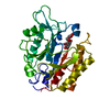
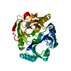
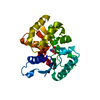
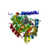
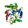
| Title | Russell's viper venom serine proteinase, RVV-V in complex with the fragment (residues 1533-1546) of human factor V | |||||||||
 Components Components |
| |||||||||
 Keywords Keywords | HYDROLASE / serine proteinase / double six-stranded beta-barrels / glycosylation | |||||||||
| Function / homology |  Function and homology information Function and homology informationsnake venom factor V activator / response to vitamin K / platelet alpha granule / Cargo concentration in the ER / COPII-coated ER to Golgi transport vesicle / COPII-mediated vesicle transport / blood circulation / : / endoplasmic reticulum-Golgi intermediate compartment membrane / secretory granule ...snake venom factor V activator / response to vitamin K / platelet alpha granule / Cargo concentration in the ER / COPII-coated ER to Golgi transport vesicle / COPII-mediated vesicle transport / blood circulation / : / endoplasmic reticulum-Golgi intermediate compartment membrane / secretory granule / platelet alpha granule lumen / Post-translational protein phosphorylation / Regulation of Insulin-like Growth Factor (IGF) transport and uptake by Insulin-like Growth Factor Binding Proteins (IGFBPs) / blood coagulation / Platelet degranulation / toxin activity / extracellular vesicle / endoplasmic reticulum lumen / copper ion binding / serine-type endopeptidase activity / proteolysis / : / extracellular region / membrane / plasma membrane Similarity search - Function | |||||||||
| Biological species |  Daboia russellii siamensis (Siamese Russell's viper) Daboia russellii siamensis (Siamese Russell's viper) Homo sapiens (human) Homo sapiens (human) | |||||||||
| Method |  X-RAY DIFFRACTION / SYNCHROTRON / MOLECULAR REPLACEMENT / Resolution: 1.8 Å X-RAY DIFFRACTION / SYNCHROTRON / MOLECULAR REPLACEMENT / Resolution: 1.8 Å | |||||||||
 Authors Authors | Nakayama, D. / Ben Ammar, Y. / Takeda, S. | |||||||||
 Citation Citation | Journal: Febs Lett. / Year: 2011 Title: Structural basis of coagulation factor V recognition for cleavage by RVV-V Authors: Nakayama, D. / Ben Ammar, Y. / Miyata, T. / Takeda, S. #1: Journal: Acta Crystallogr.,Sect.F / Year: 2009 Title: Crystallization and preliminary X-ray crystallographic analysis of blood coagulation factor V-activating proteinase (RVV-V) from Russell's viper venom Authors: Nakayama, D. / Ben Ammar, Y. / Takeda, S. | |||||||||
| History |
|
- Structure visualization
| Structure viewer | Molecule: MolmilJmol/JSmol |
|---|
- Downloads & links
-Download
| PDBx/mmCIF format | 3s9c.cif.gz | 71.3 KB | Display | PDBx/mmCIF format |
|---|---|---|---|---|
| PDB format | pdb3s9c.ent.gz | 50.1 KB | Display | PDB format |
| PDBx/mmJSON format | 3s9c.json.gz | Tree view | PDBx/mmJSON format | |
| Others |  Other downloads Other downloads |
-Validation report
| Arichive directory | https://data.pdbj.org/pub/pdb/validation_reports/s9/3s9cftp://data.pdbj.org/pub/pdb/validation_reports/s9/3s9c | HTTPS FTP |
|---|
-Related structure data
| Related structure data |  3s9aSC  3s9bC  3sbkC S: Starting model for refinement C: citing same article ( |
|---|---|
| Similar structure data |


-Links
 PDBj
PDBj


- Assembly
| Deposited unit | 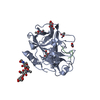
| ||||||||
|---|---|---|---|---|---|---|---|---|---|
| 1 |
| ||||||||
| Unit cell |
|
-Components
-Protein / Protein/peptide , 2 types, 2 molecules AB
| #1: Protein | Mass: 25960.971 Da / Num. of mol.: 1 / Source method: isolated from a natural source Source: (natural) Daboia russellii siamensis (Siamese Russell's viper)Tissue: venom / References: UniProt: P18965, snake venom factor V activator |
|---|---|
| #2: Protein/peptide | Mass: 1665.806 Da / Num. of mol.: 1 / Fragment: UNP residues 1561-1574 / Source method: obtained synthetically / Details: synthetic peptide / Source: (synth.) Homo sapiens (human) / References: UniProt: P12259 |
-Sugars , 3 types, 3 molecules 

| #3: Polysaccharide | beta-D-mannopyranose-(1-4)-2-acetamido-2-deoxy-beta-D-glucopyranose-(1-4)-2-acetamido-2-deoxy-beta- ...beta-D-mannopyranose-(1-4)-2-acetamido-2-deoxy-beta-D-glucopyranose-(1-4)-2-acetamido-2-deoxy-beta-D-glucopyranose Source method: isolated from a genetically manipulated source |
|---|---|
| #4: Sugar | ChemComp-BGC /  Type: D-saccharide, beta linking / Mass: 180.156 Da / Num. of mol.: 1 Type: D-saccharide, beta linking / Mass: 180.156 Da / Num. of mol.: 1Source method: isolated from a genetically manipulated source Formula: C6H12O6 |
| #5: Sugar | ChemComp-GLC /  Type: D-saccharide, alpha linking / Mass: 180.156 Da / Num. of mol.: 1 Type: D-saccharide, alpha linking / Mass: 180.156 Da / Num. of mol.: 1Source method: isolated from a genetically manipulated source Formula: C6H12O6 |
-Non-polymers , 3 types, 203 molecules 


| #6: Chemical | ChemComp-ACT /  Mass: 59.044 Da / Num. of mol.: 4 / Source method: obtained synthetically / Formula: C2H3O2 Mass: 59.044 Da / Num. of mol.: 4 / Source method: obtained synthetically / Formula: C2H3O2#7: Chemical |  Mass: 65.409 Da / Num. of mol.: 3 / Source method: obtained synthetically / Formula: Zn Mass: 65.409 Da / Num. of mol.: 3 / Source method: obtained synthetically / Formula: Zn#8: Water | ChemComp-HOH / | Mass: 18.015 Da / Num. of mol.: 196 / Source method: isolated from a natural source / Formula: H2O |
|---|
-Details
| Has protein modification | Y |
|---|---|
| Sequence details | THE RESIDUE NUMBERING IS NOT SEQUENTIAL. THE RESIDUE NUMBERING IS BASED ON THE TOPOLOGICAL ...THE RESIDUE NUMBERING IS NOT SEQUENTIAL |
-Experimental details
-Experiment
| Experiment | Method: X-RAY DIFFRACTION / Number of used crystals: 1 |
|---|
- Sample preparation
| Crystal | Density Matthews: 2.36 Å3/Da / Density % sol: 47.99 % |
|---|---|
| Crystal grow | Temperature: 293 K / Method: vapor diffusion, sitting drop / pH: 6 Details: 20% PEG 3350, 0.2M zinc acetate, pH 6.0, VAPOR DIFFUSION, SITTING DROP, temperature 293K |
-Data collection
| Diffraction | Mean temperature: 100 K |
|---|---|
| Diffraction source | Source: SYNCHROTRON / Site: SPring-8  / Beamline: BL41XU / Wavelength: 1 Å / Beamline: BL41XU / Wavelength: 1 Å |
| Detector | Type: RAYONIX MX-225 / Detector: CCD / Date: Oct 13, 2009 / Details: mirrors |
| Radiation | Monochromator: Rotated-inclined double-crystal / Protocol: SINGLE WAVELENGTH / Monochromatic (M) / Laue (L): M / Scattering type: x-ray |
| Radiation wavelength | Wavelength: 1 Å / Relative weight: 1 |
| Reflection | Resolution: 1.8→30 Å / Num. all: 24272 / Num. obs: 24029 / % possible obs: 99 % / Observed criterion σ(F): 0 / Observed criterion σ(I): 0 / Redundancy: 10.9 % / Biso Wilson estimate: 19.2 Å2 / Rmerge(I) obs: 0.047 / Net I/σ(I): 46.3 |
| Reflection shell | Resolution: 1.8→1.86 Å / Rmerge(I) obs: 0.296 / Mean I/σ(I) obs: 6.9 / Num. unique all: 2213 / % possible all: 91.8 |
- Processing
| Software |
| ||||||||||||||||||||||||||||
|---|---|---|---|---|---|---|---|---|---|---|---|---|---|---|---|---|---|---|---|---|---|---|---|---|---|---|---|---|---|
| Refinement | Method to determine structure: MOLECULAR REPLACEMENT Starting model: PDB ENTRY 3S9A Resolution: 1.8→29.21 Å / Rfactor Rfree error: 0.006 / Data cutoff high absF: 1637135.5 / Data cutoff low absF: 0 / Isotropic thermal model: RESTRAINED / Cross valid method: THROUGHOUT / σ(F): 0 / σ(I): 0 / Stereochemistry target values: Engh & Huber / Details: BULK SOLVENT MODEL USED
| ||||||||||||||||||||||||||||
| Solvent computation | Solvent model: FLAT MODEL / Bsol: 64.5738 Å2 / ksol: 0.42 e/Å3 | ||||||||||||||||||||||||||||
| Displacement parameters | Biso mean: 33.9 Å2
| ||||||||||||||||||||||||||||
| Refine analyze |
| ||||||||||||||||||||||||||||
| Refinement step | Cycle: LAST / Resolution: 1.8→29.21 Å
| ||||||||||||||||||||||||||||
| Refine LS restraints |
| ||||||||||||||||||||||||||||
| LS refinement shell | Resolution: 1.8→1.91 Å / Rfactor Rfree error: 0.018 / Total num. of bins used: 6
| ||||||||||||||||||||||||||||
| Xplor file |
|
