 Movie
Movie Controller

[English] 日本語
 Yorodumi
- PDB-1lkd: CRYSTAL STRUCTURE OF 2,3-DIHYDROXYBIPHENYL 1,2-DIOXYGENASE (DHBD)... -
+ Open data

- Basic information
| Entry | Database: PDB / ID: 1lkd | ||||||
|---|---|---|---|---|---|---|---|
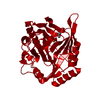
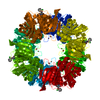
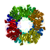
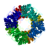
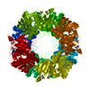
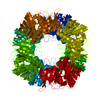
| Title | CRYSTAL STRUCTURE OF 2,3-DIHYDROXYBIPHENYL 1,2-DIOXYGENASE (DHBD) COMPLEXED WITH 2',6'-DICL DIHYDROXYBIPHENYL (DHB) | ||||||
 Components Components | BIPHENYL-2,3-DIOL 1,2-DIOXYGENASE | ||||||
 Keywords Keywords | OXIDOREDUCTASE / Extradiol dioxygenase / 2 / 3-Dihydroxybiphenyl / non-heme iron / Anaerobic / PCB biodegradation | ||||||
| Function / homology |  Function and homology information Function and homology informationbiphenyl-2,3-diol 1,2-dioxygenase / biphenyl-2,3-diol 1,2-dioxygenase activity / xenobiotic catabolic process / ferrous iron binding Similarity search - Function | ||||||
| Biological species |  Burkholderia xenovorans (bacteria) Burkholderia xenovorans (bacteria) | ||||||
| Method |  X-RAY DIFFRACTION / SYNCHROTRON / MOLECULAR REPLACEMENT / Resolution: 1.7 Å X-RAY DIFFRACTION / SYNCHROTRON / MOLECULAR REPLACEMENT / Resolution: 1.7 Å | ||||||
 Authors Authors | Dai, S. / Bolin, J.T. | ||||||
 Citation Citation | Journal: Nat.Struct.Biol. / Year: 2002 Title: Identification and analysis of a bottleneck in PCB biodegradation Authors: Dai, S. / Vaillancourt, F.H. / Maaroufi, H. / Drouin, N.M. / Neau, D.B. / Snieckus, V. / Bolin, J.T. / Eltis, L.D. | ||||||
| History |
|
- Structure visualization
| Structure viewer | Molecule: MolmilJmol/JSmol |
|---|
- Downloads & links
-Download
| PDBx/mmCIF format | 1lkd.cif.gz | 79 KB | Display | PDBx/mmCIF format |
|---|---|---|---|---|
| PDB format | pdb1lkd.ent.gz | 57.4 KB | Display | PDB format |
| PDBx/mmJSON format | 1lkd.json.gz | Tree view | PDBx/mmJSON format | |
| Others |  Other downloads Other downloads |
-Validation report
| Arichive directory | https://data.pdbj.org/pub/pdb/validation_reports/lk/1lkdftp://data.pdbj.org/pub/pdb/validation_reports/lk/1lkd | HTTPS FTP |
|---|
-Related structure data


-Links
 PDBj
PDBj


- Assembly
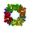
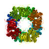
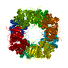
| Deposited unit | 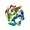
| ||||||||
|---|---|---|---|---|---|---|---|---|---|
| 1 | x 8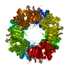
| ||||||||
| Unit cell |
| ||||||||
| Components on special symmetry positions |
|
-Components
| #1: Protein | Mass: 32377.598 Da / Num. of mol.: 1 Source method: isolated from a genetically manipulated source Source: (gene. exp.) Burkholderia xenovorans (bacteria) / Strain: LB400Description: Burkholderia sp. strain LB400 has been reclassifed. Prior publications may refer to this organism as pseudomonas sp. Strain LB400 or burkholderia cepacia strain LB400. See M.G.Fain,J.D. ...Description: Burkholderia sp. strain LB400 has been reclassifed. Prior publications may refer to this organism as pseudomonas sp. Strain LB400 or burkholderia cepacia strain LB400. See M.G.Fain,J.D.Haddock, Current Microbiol. (2001) 42:269-73 Plasmid: pLEBD4 / Production host: Pseudomonas putida (bacteria) / Strain (production host): KT2442References: UniProt: P47228, biphenyl-2,3-diol 1,2-dioxygenase | ||||||
|---|---|---|---|---|---|---|---|
| #2: Chemical |   Mass: 55.845 Da / Num. of mol.: 2 / Source method: obtained synthetically / Formula: Fe Mass: 55.845 Da / Num. of mol.: 2 / Source method: obtained synthetically / Formula: Fe#3: Chemical |   Mass: 255.097 Da / Num. of mol.: 2 / Source method: obtained synthetically / Formula: C12H8Cl2O2 Mass: 255.097 Da / Num. of mol.: 2 / Source method: obtained synthetically / Formula: C12H8Cl2O2#4: Chemical | ChemComp-P6G / |   Mass: 282.331 Da / Num. of mol.: 1 / Source method: obtained synthetically / Formula: C12H26O7 / Comment: precipitant*YM Mass: 282.331 Da / Num. of mol.: 1 / Source method: obtained synthetically / Formula: C12H26O7 / Comment: precipitant*YM#5: Water | ChemComp-HOH / |  Mass: 18.015 Da / Num. of mol.: 326 / Source method: isolated from a natural source / Formula: H2O Mass: 18.015 Da / Num. of mol.: 326 / Source method: isolated from a natural source / Formula: H2O |
-Experimental details
-Experiment
| Experiment | Method: X-RAY DIFFRACTION / Number of used crystals: 1 |
|---|
- Sample preparation
| Crystal | Density Matthews: 3.1 Å3/Da / Density % sol: 60 % | |||||||||||||||
|---|---|---|---|---|---|---|---|---|---|---|---|---|---|---|---|---|
| Crystal grow | Temperature: 283 K / Method: vapor diffusion, sitting drop / pH: 7.5 Details: PEG 400, t-Butanol, pH 7.5, VAPOR DIFFUSION, SITTING DROP, temperature 283K | |||||||||||||||
| Crystal grow | *PLUS Temperature: 5-10 ℃ / Method: vapor diffusion / Details: Han, S., (1995) Science, 270, 976. | |||||||||||||||
| Components of the solutions | *PLUS
|
-Data collection
| Diffraction | Mean temperature: 100 K |
|---|---|
| Diffraction source | Source: SYNCHROTRON / Site: APS  / Beamline: 19-ID / Wavelength: 0.97833 Å / Beamline: 19-ID / Wavelength: 0.97833 Å |
| Detector | Type: CUSTOM-MADE / Detector: CCD / Date: Jun 10, 2001 |
| Radiation | Monochromator: SAGITALLY FOCUSED Si (III) / Protocol: SINGLE WAVELENGTH / Monochromatic (M) / Laue (L): M / Scattering type: x-ray |
| Radiation wavelength | Wavelength: 0.97833 Å / Relative weight: 1 |
| Reflection | Resolution: 1.69→50 Å / Num. all: 46029 / Num. obs: 46029 / % possible obs: 90.2 % / Observed criterion σ(F): 0 / Observed criterion σ(I): -3 / Biso Wilson estimate: 16.9 Å2 / Rmerge(I) obs: 0.086 |
| Reflection shell | Resolution: 1.69→1.75 Å / Rmerge(I) obs: 0.409 / % possible all: 73.7 |
| Reflection | *PLUS Num. measured all: 2212442 |
| Reflection shell | *PLUS % possible obs: 73.7 % |
- Processing
| Software |
| ||||||||||||||||||||||||||||||||||||
|---|---|---|---|---|---|---|---|---|---|---|---|---|---|---|---|---|---|---|---|---|---|---|---|---|---|---|---|---|---|---|---|---|---|---|---|---|---|
| Refinement | Method to determine structure: MOLECULAR REPLACEMENT / Resolution: 1.7→19.97 Å / Rfactor Rfree error: 0.005 / Data cutoff high absF: 2808076.19 / Data cutoff high rms absF: 2808076.19 / Data cutoff low absF: 0 / Isotropic thermal model: RESTRAINED / Cross valid method: THROUGHOUT / σ(F): 0 / Stereochemistry target values: Engh & Huber
| ||||||||||||||||||||||||||||||||||||
| Solvent computation | Solvent model: FLAT MODEL / Bsol: 61.3977 Å2 / ksol: 0.413408 e/Å3 | ||||||||||||||||||||||||||||||||||||
| Displacement parameters | Biso mean: 22.6 Å2
| ||||||||||||||||||||||||||||||||||||
| Refine analyze | Luzzati coordinate error free: 0.24 Å / Luzzati sigma a free: 0.2 Å | ||||||||||||||||||||||||||||||||||||
| Refinement step | Cycle: LAST / Resolution: 1.7→19.97 Å
| ||||||||||||||||||||||||||||||||||||
| Refine LS restraints |
| ||||||||||||||||||||||||||||||||||||
| LS refinement shell | Resolution: 1.7→1.81 Å / Rfactor Rfree error: 0.021 / Total num. of bins used: 6
| ||||||||||||||||||||||||||||||||||||
| Xplor file |
| ||||||||||||||||||||||||||||||||||||
| Refinement | *PLUS Highest resolution: 1.7 Å / Lowest resolution: 25 Å / Rfactor Rfree: 0.231 / Rfactor Rwork: 0.212 | ||||||||||||||||||||||||||||||||||||
| Solvent computation | *PLUS | ||||||||||||||||||||||||||||||||||||
| Displacement parameters | *PLUS | ||||||||||||||||||||||||||||||||||||
| Refine LS restraints | *PLUS
|
