 Movie
Movie Controller

[English] 日本語
 Yorodumi
- PDB-1knf: Crystal Structure of 2,3-dihydroxybiphenyl 1,2-dioxygenase Comple... -
+ Open data

- Basic information
| Entry | Database: PDB / ID: 1knf | ||||||
|---|---|---|---|---|---|---|---|
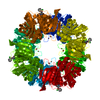
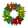
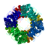
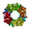
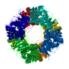
| Title | Crystal Structure of 2,3-dihydroxybiphenyl 1,2-dioxygenase Complexed with 3-methyl Catechol under Anaerobic Condition | ||||||
 Components Components | 2,3-DIHYDROXYBIPHENYL 1,2-DIOXYGENASE | ||||||
 Keywords Keywords | OXIDOREDUCTASE / dioxygenase / 2 / 3-dihydroxybiphenyl / 3-methyl catechol | ||||||
| Function / homology |  Function and homology information Function and homology informationbiphenyl-2,3-diol 1,2-dioxygenase / biphenyl-2,3-diol 1,2-dioxygenase activity / xenobiotic catabolic process / ferrous iron binding Similarity search - Function | ||||||
| Biological species |  Burkholderia xenovorans (bacteria) Burkholderia xenovorans (bacteria) | ||||||
| Method |  X-RAY DIFFRACTION / MOLECULAR REPLACEMENT / Resolution: 1.9 Å X-RAY DIFFRACTION / MOLECULAR REPLACEMENT / Resolution: 1.9 Å | ||||||
 Authors Authors | Han, S. / Bolin, J.T. | ||||||
 Citation Citation | Journal: J.Biol.Chem. / Year: 1998 Title: Molecular basis for the stabilization and inhibition of 2, 3-dihydroxybiphenyl 1,2-dioxygenase by t-butanol. Authors: Vaillancourt, F.H. / Han, S. / Fortin, P.D. / Bolin, J.T. / Eltis, L.D. #1: Journal: Science / Year: 1995Title: Crystal Structure of the Biphenyl-cleaving Extradiol Dioxygenase from a PCB-degrading Pseudomonad. Authors: Han, S. / Eltis, L.D. / Timmis, K.N. / Muchmore, S.W. / Bolin, J.T. #2: Journal: Handbook of Metalloproteins / Year: 2001Title: 2,3-Dihydroxybiphenyl 1,2-dioxygenase. Authors: Bolin, J.T. / Eltis, L.D. | ||||||
| History |
|
- Structure visualization
| Structure viewer | Molecule: MolmilJmol/JSmol |
|---|
- Downloads & links
-Download
| PDBx/mmCIF format | 1knf.cif.gz | 74.2 KB | Display | PDBx/mmCIF format |
|---|---|---|---|---|
| PDB format | pdb1knf.ent.gz | 53.8 KB | Display | PDB format |
| PDBx/mmJSON format | 1knf.json.gz | Tree view | PDBx/mmJSON format | |
| Others |  Other downloads Other downloads |
-Validation report
| Arichive directory | https://data.pdbj.org/pub/pdb/validation_reports/kn/1knfftp://data.pdbj.org/pub/pdb/validation_reports/kn/1knf | HTTPS FTP |
|---|
-Related structure data
| Related structure data | 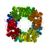 1kmyC 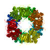 1kndC 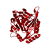 1hanS S: Starting model for refinement C: citing same article ( |
|---|---|
| Similar structure data |


-Links
 PDBj
PDBj


- Assembly
| Deposited unit | 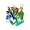
| ||||||||
|---|---|---|---|---|---|---|---|---|---|
| 1 | x 8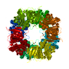
| ||||||||
| Unit cell |
| ||||||||
| Details | The biological assembly is homo-octamer generated by crystallographic symmetry |
-Components
| #1: Protein | Mass: 32377.598 Da / Num. of mol.: 1 Source method: isolated from a genetically manipulated source Source: (gene. exp.) Burkholderia xenovorans (bacteria) / Strain: LB400Description: HYPEREXPRESSED IN THE PARENT STRAIN (This organism has been reclassified. Prior publications may refer to this source as Pseudomonas sp. strain LB400.) Gene: BPHC / Plasmid: PLEBD4 / Production host: Burkholderia cepacia (bacteria)References: UniProt: P47228, biphenyl-2,3-diol 1,2-dioxygenase | ||||||
|---|---|---|---|---|---|---|---|
| #2: Chemical |   Mass: 55.845 Da / Num. of mol.: 2 / Source method: obtained synthetically / Formula: Fe Mass: 55.845 Da / Num. of mol.: 2 / Source method: obtained synthetically / Formula: Fe#3: Chemical | ChemComp-MBD / |   Mass: 124.137 Da / Num. of mol.: 1 / Source method: obtained synthetically / Formula: C7H8O2 Mass: 124.137 Da / Num. of mol.: 1 / Source method: obtained synthetically / Formula: C7H8O2#4: Chemical |   Mass: 74.122 Da / Num. of mol.: 2 / Source method: obtained synthetically / Formula: C4H10O Mass: 74.122 Da / Num. of mol.: 2 / Source method: obtained synthetically / Formula: C4H10O#5: Water | ChemComp-HOH / |  Mass: 18.015 Da / Num. of mol.: 121 / Source method: isolated from a natural source / Formula: H2O Mass: 18.015 Da / Num. of mol.: 121 / Source method: isolated from a natural source / Formula: H2O |
-Experimental details
-Experiment
| Experiment | Method: X-RAY DIFFRACTION / Number of used crystals: 1 |
|---|
- Sample preparation
| Crystal | Density Matthews: 3.22 Å3/Da / Density % sol: 62 % |
|---|---|
| Crystal grow | Temperature: 278 K / Method: vapor diffusion, sitting drop / pH: 7.5 Details: PEG4000, t-butanol, 3-methyl catechol, pH 7.5, VAPOR DIFFUSION, SITTING DROP, temperature 278K |
-Data collection
| Diffraction | Mean temperature: 298 K |
|---|---|
| Diffraction source | Source: ROTATING ANODE / Type: RIGAKU RU200 / Wavelength: 1.5418 Å |
| Detector | Type: RIGAKU RAXIS II / Detector: IMAGE PLATE / Date: Nov 30, 1995 / Details: mirrors |
| Radiation | Monochromator: YALE MIRRORS / Protocol: SINGLE WAVELENGTH / Monochromatic (M) / Laue (L): M / Scattering type: x-ray |
| Radiation wavelength | Wavelength: 1.5418 Å / Relative weight: 1 |
| Reflection | Resolution: 1.9→40 Å / Num. all: 32641 / Num. obs: 32641 / % possible obs: 97 % / Observed criterion σ(F): 0 / Observed criterion σ(I): 0 / Redundancy: 6.9 % / Rsym value: 0.08 / Net I/σ(I): 44.8 |
| Reflection shell | Resolution: 1.9→1.97 Å / Redundancy: 3.8 % / Mean I/σ(I) obs: 5.1 / Num. unique all: 2397 / Rsym value: 0.27 / % possible all: 73.1 |
- Processing
| Software |
| ||||||||||||||||||||||||||||
|---|---|---|---|---|---|---|---|---|---|---|---|---|---|---|---|---|---|---|---|---|---|---|---|---|---|---|---|---|---|
| Refinement | Method to determine structure: MOLECULAR REPLACEMENT Starting model: 1HAN Resolution: 1.9→7 Å / Cross valid method: THROUGHOUT / σ(F): 0 / σ(I): 0 / Stereochemistry target values: Engh & Huber Details: ALL FE-PROTEIN BOND DISTANCES WERE HARMONICALLY RESTRAINED TO AN EQUILIBRIUM DISTANCE OF 2.2 ANGSTROMS USING A WEAK FORCE CONSTANT OF 10 KCAL/(MOLE X ANGSTROM-SQUARED). BOND LENGTH, BOND ...Details: ALL FE-PROTEIN BOND DISTANCES WERE HARMONICALLY RESTRAINED TO AN EQUILIBRIUM DISTANCE OF 2.2 ANGSTROMS USING A WEAK FORCE CONSTANT OF 10 KCAL/(MOLE X ANGSTROM-SQUARED). BOND LENGTH, BOND ANGLE, AND PLANARITY RESTRAINTS SIMILAR TO THOSE USED FOR AROMATIC SIDE CHAINS WERE APPLIED TO HET GROUP CAQ (CATECHOL). FE-CAQ AND FE-WATER BOND DISTANCES WERE NOT RESTRAINED. THE REFINED MODEL INCLUDES TWO MUTALLY EXCLUSIVE STRUCTURES IN THE VICINITY OF THE ACTIVE SITE. STRUCTURE ONE (labeled with alternate conformation marker A) INCLUDES A SUBSTRATE 3-METHYL-CATECHOL (MBD 301) AND WATERS 9001 and 9002 AT 50% OCCUPANCY. ATOMS OA1 AND OA2 OF MBD 301 AND WATER 9001 ARE COORDINATED TO FE2 500. STRUCTURE TWO (labeled with alternate conformation marker B) INCLUDES ONE MOLECULE OF T-BUTANOL (TBU 600) AND WATERS 3001, 3012, AND 4014 AT 50% OCCUPANCY, AND IS EQUIVALENT TO THE SUBSTRATE-FREE STRUCTURE WHERE WATERS 3001 AND 3012 ARE COORDINATED TO FE2 500.
| ||||||||||||||||||||||||||||
| Displacement parameters | Biso mean: 24.3 Å2 | ||||||||||||||||||||||||||||
| Refinement step | Cycle: LAST / Resolution: 1.9→7 Å
| ||||||||||||||||||||||||||||
| Refine LS restraints |
| ||||||||||||||||||||||||||||
| LS refinement shell | Resolution: 1.9→1.93 Å
|
